Pathology Image Detail
Caption |
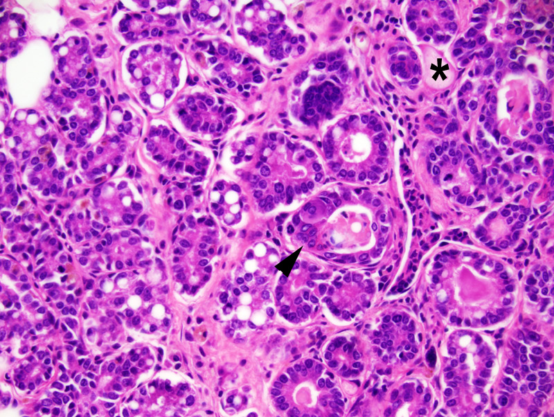
Hyperplasia and squamous metaplasia, mammary gland: this photomicrograph illustrates an area of transition between an area of hyperplasia of the mammary gland (on the left) and an area of squamous metaplasia (on the right). A raft of ghost epithelial cells (*) and multinucleated epithelial cells (arrowhead) are present. A few nuclei have an apical location instead of a basal location, a feature consistent with dysplasia. |
Description |
Hyperplasia, ductal and alveolar, with squamous metaplasia, mammary gland |
Age at Necropsy |
unknown |
Contributor |
Ward JM (J:107304) |
Pathologist |
Mikaelian I (J:94320) |
Method |
H&E |
Model |
|
Strain |
|