Caption |
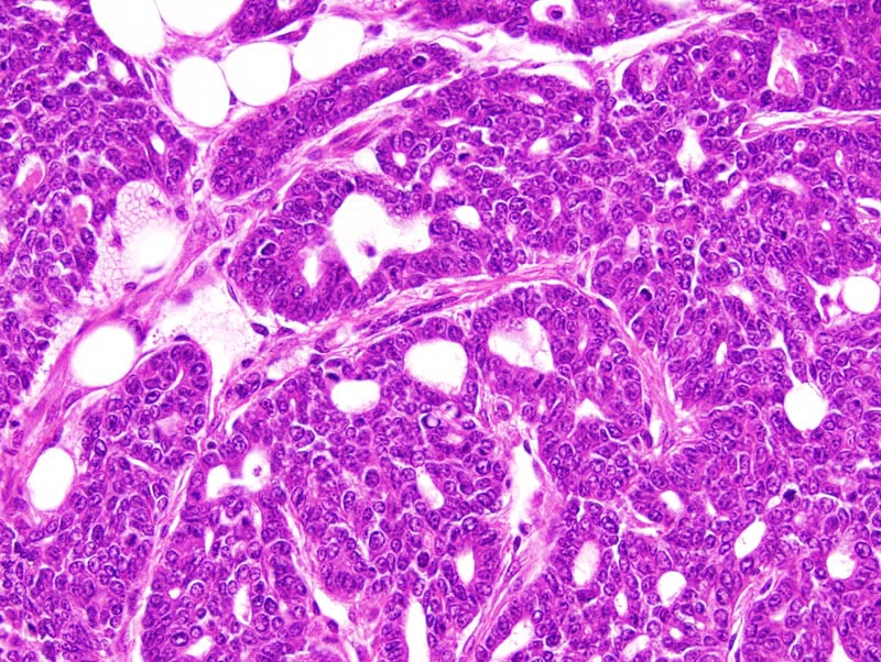
Mammary gland: this photomicrograph focuses on the periphery of the neoplasm where glandular differentiation is prominent. A few adipocytes are entrapped within the neoplasm. The glands are lined by a one cell-thick cuboidal epithelium where neoplastic cells often pile-up disorderly. Neoplastic cells have ill-defined cell borders and a moderate amount of acidophilic cytoplasm. The nucleus is central, oval, slightly hypochromatic, with a coarsely clumped chromatin and 1-3 small basophilic nucleoli. Anisokaryosis and anisocytosis are mild. Mitoses are numerous. |
Description |
Adenocarcinoma, solid, glandular, mammary gland |
Age at Necropsy |
unknown |
Notes |
This neoplasm illustrates the most common phenotype of neoplasms arising in mice transgenic for Notch4. Smaller neoplasms tend to be predominantly composed of glandular areas while larger neoplasms generally are predominantly composed of solid areas. However, glandular and solid areas are present in most mammary tumors arising in mice transgenic for Notch4. |
Contributor |
Ward JM (J:107304) |
Pathologist |
Mikaelian I (J:94320) |
Method |
H&E |