Pathology Image Detail
Caption |
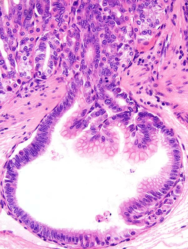
Stomach: this high magnification photomicrograph represents gastric gland herniated underneath the muscularis mucosae. Cells lining the herniated gland do not show atypia or a high mitotic rate. These cells have undergone goblet cell differentiation (catarrhal metaplasia). The herniated gland is not surrounded by a scirrhous reaction. |
Description |
Cystic hyperplasia, glandular stomach |
Age at Necropsy |
305 days |
Contributor |
Churchill GA (J:112750) |
Pathologist |
Mikaelian I (J:94320) |
Method |
H&E |
Model |
|
Strain |
|