Pathology Image Detail

Caption

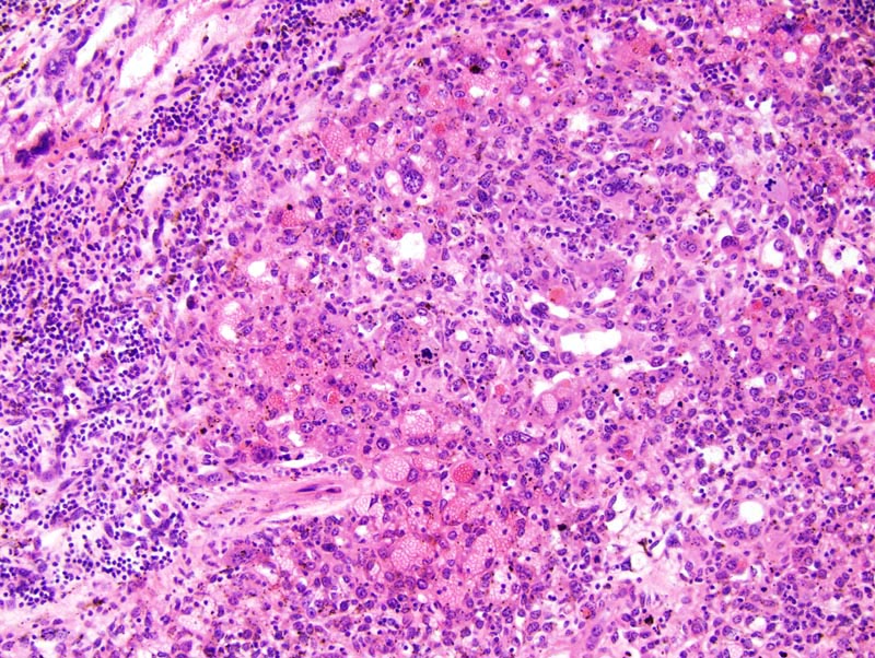
Subcutis: the subcutis is expanded by an ill defined, unencapsulated, invasive, multilobular, densely cellular neoplasm which contains large pools of blood. The neoplasm is composed of solid areas and interconnected and tortuous blood-filed vascular spaces supported by a scant amount of fibrovascular stroma. Neoplastic cells in this portion of the neoplasm are medium-sized, histiocytoid, with indistinct cell borders and with a moderate amount of strongly acidophilic cytoplasm. The nucleus is central, round to oval, normochromatic, with a clumped chromatin. There is prominent lymphocytic infiltration at the periphery of the tumor. The tumor is infiltrated by a moderate number of hemosiderin-ladden macrophages.

Description

Hemangiosarcoma, subcutis

Age at Necropsy

50 days

Contributor

Mikaelian I (J:94320)

Pathologist

Mikaelian I (J:94320)

Method

H&E

Model

MMHC ID
MTB:29451
Tumor Name
Blood vessel • hemangiosarcoma
Treatment Type
None (spontaneous)
Tumor Synonyms
Hemangiosarcoma, subcutis
Organ Affected
Subcutis
Frequency
observed
Reference
J:94320

Strain

Strain
B6.129S2-Trp53tm1Tyj/J
Strain Types
congenic
targeted mutation (knockout)
General Note
Heterozygous mice. The genetic background of these mice is congenic C57BL/6J.
Strain Synonyms
B6.129S2-Trp53tm1Tyj
C57BL/6J-Trp53tm1Tyj
p53+/-
p53 heterozygous (+/-)
C57BL/6J-Trp53tm1Tyj
p53(+/-)
B6.129S2-Trp53tm1Tyj
p53+/-
p53+/-
Strain Sex
Male
Reproductive Status
reproductive status not specified
Age of Detection
50 days