Pathology Image Detail
Caption |
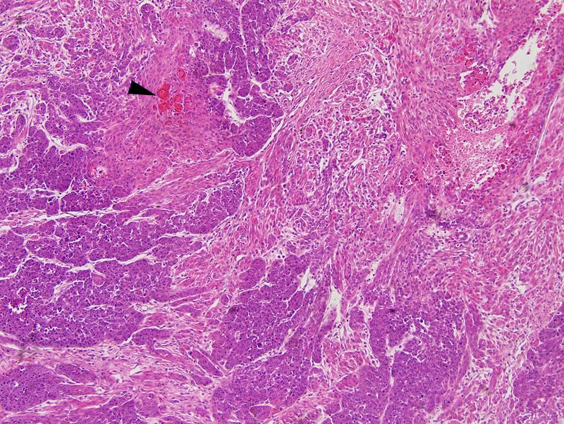
Mammary gland: this photomicrograph illustrates an areas of epithelial to mesenchymal transition. The portions of the photomicrograph on the left and in the lower portions of the photomicrograph correspond to solid portions of the neoplasm. The upper right corner of the photomicrograph represents a mesenchymal portion of the neoplasm. A focus of cornification (arrowhead) is present at the center of a large solid area. |
Description |
Carcinoma, solid, comedo, with squamous differentiation and epithelial to mesenchymal transition, mammary gland |
Age at Necropsy |
250 days |
Contributor |
Churchill GA (J:112750) |
Pathologist |
Mikaelian I (J:94320) |
Method |
H&E |
Model |
|
Strain |
|