Pathology Image Detail
Caption |
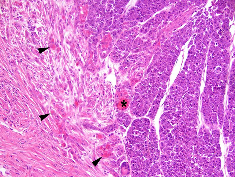
Mammary gland: this photomicrograph illustrates an area of epithelial to mesenchymal transition. There is gradual transition between cords and solid areas of neoplastic cells with an epithelial phenotype to bundles of neoplastic cells with a spindloid (= mesenchymal) phenotype. Clusters of neoplastic cells undergo cornification and form "keratin pearls" (*). There is also cornification of individual neoplastic cells (arrowheads). |
Description |
Carcinoma, solid, comedo, with squamous differentiation and epithelial to mesenchymal transition, mammary gland |
Age at Necropsy |
250 days |
Contributor |
Churchill GA (J:112750) |
Pathologist |
Mikaelian I (J:94320) |
Method |
H&E |
Model |
|
Strain |
|