Caption |
This image was submitted by JM Ward. Image is from study set created by Herbert C. Morse III (NIAID, NIH), Torgny Fredrickson (NIAID, NIH), and Jerrold M. Ward (NCI, NIH) in 2001. A whole-slide scan image cane be viewed at https://images.jax.org/webclient/img_detail/15587. |
Description |
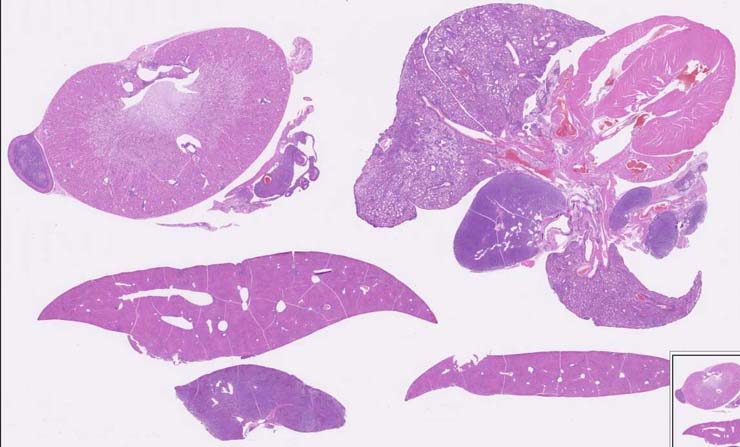
histiocyte-rich lymphoma |
Notes |
Diffuse large B-cell lymphoma, centroblastic, histiocyte-rich, spleen, lymph nodes. Most of the spleen and mediastinal lymph nodes are infiltrated with a mixture of immature lymphocytes and histiocytes. Little red pulp remains. The immature lymphoid cells (centroblasts) are mostly round in shape, possess little cytoplasm and some have a prominent nucleolus. Interspersed among the lymphoid tumor cells are large histiocytic cells with prominent eosinophilic cytoplasm. Foci of this mixed population are seen in the liver. The lung parenchyma contains the same mixed lymphoid population. Tumor cells were CD45R+. CD3+ infiltrating non-neoplastic lymphocytes were also seen. Repoterd CD45R and CD3 staining patterns have no submitted images. |
Contributor |
Ward JM (J:107304) |
Pathologist |
Ward JM (J:107304) |
Copyright |
This work was supported in part with funds from the Mouse Model of Cancers Consortium, National Cancer Institute, National Institutes of health, and by contract N01-C056000 from the National cancer Institute. |
Method |
H&E |