Caption |
This image was submitted by JM Ward. Image is from study set created by Herbert C. Morse III (NIAID, NIH), Torgny Fredrickson (NIAID, NIH), and Jerrold M. Ward (NCI, NIH) in 2001. A whole-slide scan image cane be viewed at https://images.jax.org/webclient/img_detail/15635. |
Description |
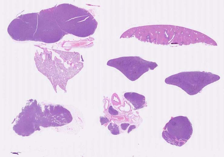
Myeloid leukemia, undifferentiated, without maturation, spleen |
Notes |
Myeloid leukemia, undifferentiated, without maturation, spleen, liver, lymph node. In the spleen, there are nodules of undifferentiated tumor cells in the red pulp and in some PALS areas. Apoptotic tumor cells are scattered among the tumor nodules. Erythroid hyperplasia is noted in the red pulp. Atrophy of the white pulp is observed. The mediastinal lymph node is infiltrated by tumor cells. Tumor cell foci are seen in liver. Foci of better differentiated myeloid cells associated with immature myeloid cells are seen that appear to represent tumor cell differentiation rather than a separate myeloid hyperplasia. The tumor cells were CD45R- and CD3-. |
Contributor |
Ward JM (J:107304) |
Pathologist |
Ward JM (J:107304) |
Copyright |
This work was supported in part with funds from the Mouse Model of Cancers Consortium, National Cancer Institute, National Institutes of health, and by contract N01-C056000 from the National cancer Institute. |
Method |
H&E |