Caption |
This image was submitted by JM Ward. Image is from study set created by Herbert C. Morse III (NIAID, NIH), Torgny Fredrickson (NIAID, NIH), and Jerrold M. Ward (NCI, NIH) in 2001. This slide was immunostained for CD3. A whole-slide scan of this image can be viewed at https://images.jax.org/webclient/img_detail/15566. |
Description |
follicular center cell lymphoma |
Notes |
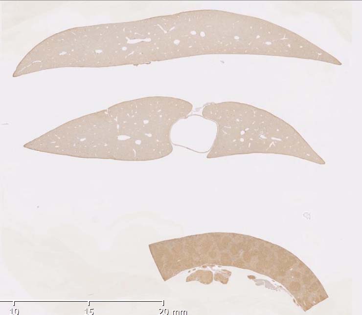
Follicular lymphoma, with focal histiocytic sarcoma, spleen. The normal structure of the white pulp cannot be seen. The white pulp areas are enlarged and infiltrated with a pleomorphic population of lymphoid cells. Centroblasts, centrocytes and small lymphocytes are seen. There is one focus of histiocytic sarcoma, with giant cells. The lymphoma cells are CD45R+ and non-neoplastic infiltrating small lymphocytes are CD3+. No slide for CD45R staining is presented, pattern is identical as shown for Case # 00000092-8A shown in pathology record 6651. |
Contributor |
Ward JM (J:107304) |
Pathologist |
Ward JM (J:107304) |
Copyright |
This work was supported in part with funds from the Mouse Model of Cancers Consortium, National Cancer Institute, National Institutes of health, and by contract N01-C056000 from the National cancer Institute. |