Caption |
This image was submitted by JM Ward. Image is from study set created by Herbert C. Morse III (NIAID, NIH), Torgny Fredrickson (NIAID, NIH), and Jerrold M. Ward (NCI, NIH) in 2001. This slide was immunostained for CD68. A whole-slide scan of this image can be viewed at https://images.jax.org/webclient/img_detail/15869/. |
Description |
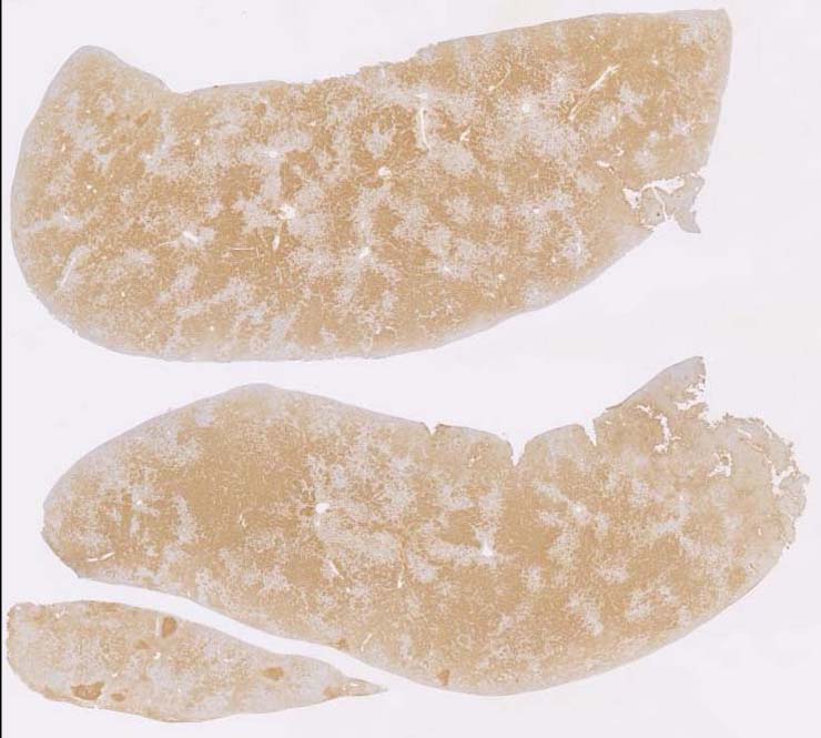
histiocytic sarcoma |
Notes |
Histiocytic sarcoma, liver. There is massive infiltration in the liver by histiocytic tumor cells. The tumor cells form vascular thrombi and emboli, have oval shaped nuclei and have abundant pale eosinophilic cytoplasm. In a few foci, the tumor cells have a spindle shape. Some multinucleated giant tumor cells are seen. Tumor cells are CD68+ and F4/80+. |
Contributor |
Ward JM (J:107304) |
Pathologist |
Ward JM (J:107304) |
Copyright |
This work was supported in part with funds from the Mouse Model of Cancers Consortium, National Cancer Institute, National Institutes of health, and by contract N01-C056000 from the National cancer Institute. |