Caption |
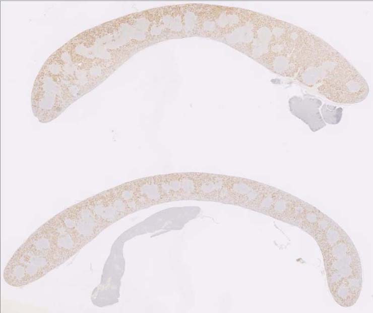
Image was submitted by Jerrald Ward. This slide was stained for INOS. Image is from study set created by Herbert C. Morse III (NIAID, NIH), Torgny Fredrickson (NIAID, NIH), and Jerrold M. Ward (NCI, NIH) in 2001. Whole-slide scan of this image is available at https://images.jax.org/webclient/img_detail/15875. |
Description |
Myeloid leukemia, preleukemic, spleen |
Notes |
Myeloid leukemia, preleukemic, spleen; Foci of immature myeloid cells can be seen beneath the splenic capsule and along splenic trabeculae. The immature cells include large immature myeloblasts with large nucleoli, ring forms (donuts), bands, and lobulated cells). They do not appear to be the normal myelopoeisis found in the spleen of mice and these lesions progress to full blown myeloid leukemia. There are more immature myeloid cells than normally seen in commonly observed myeloid hyperplasia in mice. Megakaryocyte hyperplasia associated with these myeloid cells is also noted. The white pulp is normal with some germinal centers. Bouin?s fixation. INOS IHC shows + preleukemic myeloid cells in the red pulp beneath the capsule and in the red parenchyma. |
Contributor |
Ward JM (J:107304) |
Pathologist |
Ward JM (J:107304) |
Copyright |
This work was supported in part with funds from the Mouse Model of Cancers Consortium, National Cancer Institute, National Institutes of health, and by contract N01-C056000 from the National cancer Institute. |