Caption |
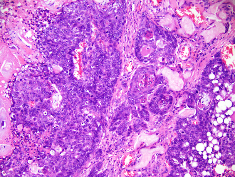
Secretory mammary adenocarcinoma with squamous differentiation: neoplastic cells, in this portion of the neoplasm, form trabeculae, nests and solid areas with central cornification. Neoplastic cells in the lower right corner of the photomicrograph contain lipid droplets. Cornification is prominent at the center of the photomicrograph. There is also a moderate granulomatous inflammation centered on rafts of "ghost" epithelial cells. |
Description |
Adenocarcinoma, secretory, with squamous differentiation, mammary gland |
Age at Necropsy |
251 days |
Notes |
This mouse had multiple mammary tumors. Each of these tumors is presented in a different pathology record. These tumors were all secretory adenocarcinomas with prominent squamous differentiation, although the relative ratio of glandular areas and areas with squamous differentiation differed among these tumors. This mouse had undergone 4 cycles of gestation and lactation. |
Contributor |
Gallego MI (J:94332) |
Pathologist |
Mikaelian I (J:94320) |
Method |
H&E |