Caption |
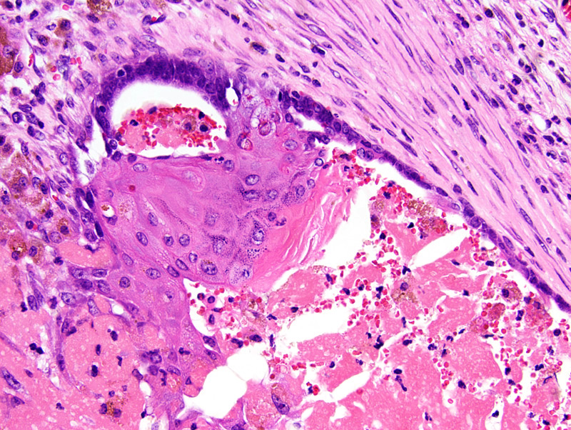
Mammary secretory adenocarcinoma: this photomicrograph illustrates an area of cornification. The neoplastic epithelium is 2-10 cells-thick and shows prominent squamous differentiation. Squamous differentiation is associated with the presence of keratohyalin granules. The epithelium surrounds a cystic space filled with degenerated neoplastic cells, blood, and fibrin. The capsule of the neoplasm is infiltrated by a moderate number of hemosiderin-laden macrophages, lymphocytes and plasma cells. As exemplified in this photomicrograph, transition from a glandular to a squamous epithelium may be abrupt. |
Description |
Adenocarcinoma, secretory, with squamous differentiation, mammary gland |
Age at Necropsy |
251 days |
Notes |
This mouse had multiple mammary tumors. Each of these tumors is presented in a different pathology record. These tumors were all secretory adenocarcinomas with prominent squamous differentiation, although the relative ratio of glandular areas and areas with squamous differentiation differed among these tumors. This mouse had undergone 4 cycles of gestation and lactation. |
Contributor |
Gallego MI (J:94332) |
Pathologist |
Mikaelian I (J:94320) |
Method |
H&E |