Pathology Image Detail
Caption |
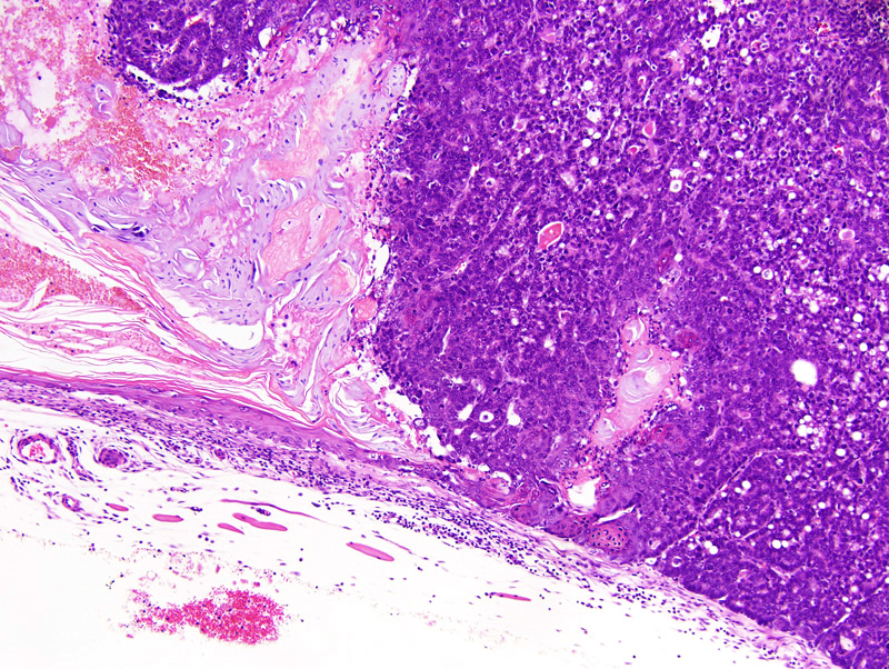
Secretory mammary adenocarcinoma with squamous differentiation: this photomicrograph illustrates an area at the junction of the cornified epithelium and the glandular portions of the neoplasm. The neoplastic cornified epithelium resembles the epidermis. The glandular portions of the neoplasm are composed of glands and solid areas supported by a scant amount of fibrovascular stroma. A few neoplastic cells contain prominent lipid droplets. |
Description |
Adenocarcinoma, secretory, with squamous differentiation, mammary gland |
Age at Necropsy |
273 days |
Notes |
This mouse had undergone 4 cycles of gestation and lactation. |
Contributor |
Gallego MI (J:94332) |
Pathologist |
Mikaelian I (J:94320) |
Method |
H&E |
Model |
|
Strain |
|