Pathology Image Detail
Caption |
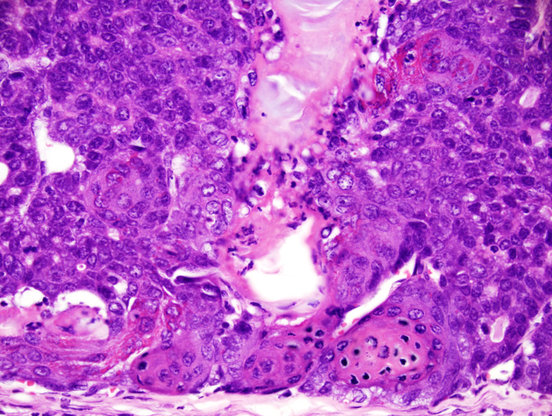
Secretory mammary adenocarcinoma with squamous differentiation: this photomicrograph illustrates an area at the junction of the cornified epithelium and the glandular and solid portions of the neoplasm. The glandular portions become solid at the vicinity of the areas of cornification. Cornification is abrupt and results in the formation of rafts of ghost epithelial cells, a feature also observed in pilomatricomas. |
Description |
Adenocarcinoma, secretory, with squamous differentiation, mammary gland |
Age at Necropsy |
273 days |
Notes |
This mouse had undergone 4 cycles of gestation and lactation. |
Contributor |
Gallego MI (J:94332) |
Pathologist |
Mikaelian I (J:94320) |
Method |
H&E |
Model |
|
Strain |
|