Pathology Image Detail
Caption |
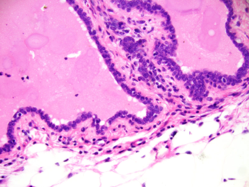
Mammary gland: this photomicrograph is centered on an interlobular mammary duct which is markedly ectatic and contains an inspissated proteinaceous fluid. The epithelium has a crowded appearance and has an increased mitotic rate compared to a normal ductal epithelium. However, epithelial cells do not pile-up. There is mild perialveolar and periductal fibrosis and pleocellular inflammation. |
Description |
Hyperplasia and ductal ectasia, mammary gland |
Age at Necropsy |
254 days |
Notes |
The mouse had undergone four cycles of gestation/lactation. |
Contributor |
Gallego MI (J:94332) |
Pathologist |
Mikaelian I (J:94320) |
Method |
H&E |
Model |
|
Strain |
|