Pathology Image Detail
Caption |
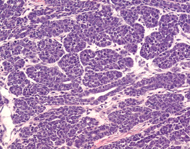
This higher magnification allows to visualize areas of glandular differentiation, as well as the formation of small solid nests of neoplastic cells. A few tubules are formed and are lined by a two-cells thick epithelium. Neoplastic cells are cuboidal to polygonal, medium-sized, with indistinct cell borders and a moderate amount of amphophilic cytoplasm. The nucleus is central, round, hyperchromatic, and with a clumped chromatin. |
Description |
Mammary gland, adenocarcinoma, acinar, solid, glandular, tubular |
Age at Necropsy |
261 days |
Contributor |
Mikaelian I (J:94320) |
Pathologist |
Mikaelian I (J:94320) |
Method |
H&E |
Model |
|
Strain |
|