Pathology Image Detail
Caption |
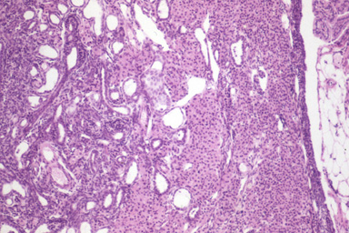
Most of the ovary is replaced by tubules resembling the surface epithelium of the ovary (left side of the figure and tubules). However, in addition, there was a thecoma composed of solid areas (right side of the figure). Neoplastic cells in these areas were polygonal, with indistinct cell borders and a large amount of delicately granular acidophilic cytoplasm. Atypia and mitotic index are minimal in both neoplasms. |
Description |
Ovary, surface epithelium adenoma and thecoma |
Age at Necropsy |
503 days |
Contributor |
Mikaelian I (J:94320) |
Pathologist |
Mikaelian I (J:94320) |
Method |
H&E |
Model |
|
Strain |
|