Pathology Image Detail

Caption

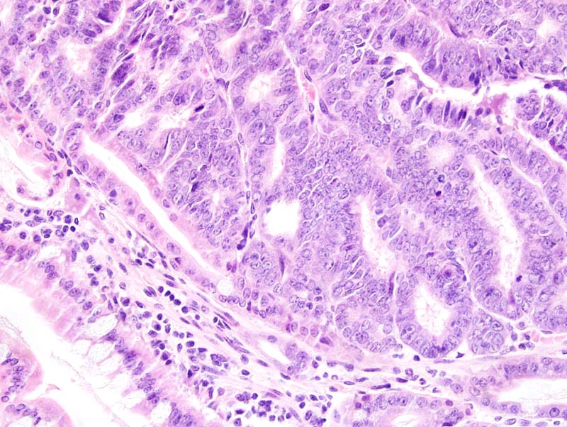
Small intestine: this high power photomicrograph illustrates the difference between the normal intestinal epithelium (lower left corner of the photomicrograph) and an intestinal adenoma. The adenoma is composed of glands lined by a one cell-thick columnar epithelium supported by a small amount of fibrovascular stroma. Neoplastic cells are columnar, with indistinct cell borders and a moderate amount of amphophilic cytoplasm. Neoplastic cells do not contain mucin droplets. Mitoses are numerous. There is no evidence of invasion in the stroma or in the submucosa.

Description

Adenoma, small intestine

Age at Necropsy

63 days

Contributor

Kemp CJ (J:97126)

Pathologist

Mikaelian I (J:94320)

Method

H&E

Model

MMHC ID
MTB:25617
Tumor Name
Intestine - Small Intestine • adenoma
Treatment Type
None (spontaneous)
Tumor Synonyms
small intestine adenoma
Organ Affected
Intestine - Small Intestine
Frequency
observed
Reference
J:94320

Strain

Strain
C57BL/6-ApcMin/+ Cdkn1btm1Mlf
Strain Types
chemically induced mutation
targeted mutation (knockout)
General Note
Mice were heterozygous for the ApcMin allele and homozygous for the Cdkn1btm1Mlf allele. These mice are the product of an intercross between strains on a predominantly C57BL/6 background.
Strain Synonyms
Min/+ p27-/-
B6.Cg-ApcMin/+ Cdkn1btm1Mlf
Strain Sex
Female
Reproductive Status
reproductive status not specified
Age of Detection
63 days