Pathology Image Detail
Caption |
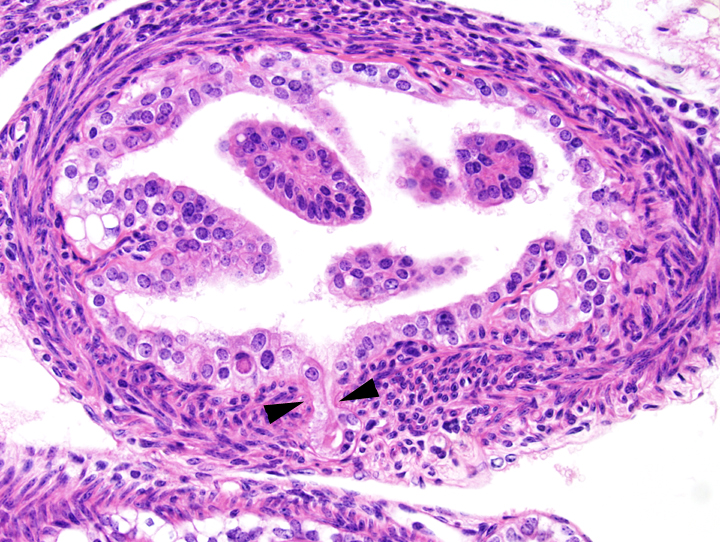
Oviduct: this photomicrograph represents the early stages of hyperplasia of the epithelium of the oviduct. There is focal herniation of the epithelium of the oviduct underneath the wall of the oviduct (arrowheads). The epithelium of the oviduct does not show evidences of atypia. There is mild multifocal vacuolation of the epithelium of the oviduct. This change is associated with a mild multifocal lymphocytic and plasmacytic infiltration of the submucosa of the oviduct. |
Description |
Hyperplasia, epithelial, mild, focal, oviduct (salpingitis isthmica nodosa) |
Age at Necropsy |
18.9 months |
Contributor |
Mikaelian I (J:94320) |
Pathologist |
Mikaelian I (J:94320) |
Method |
H&E |
Model |
|
Strain |
|