Caption |
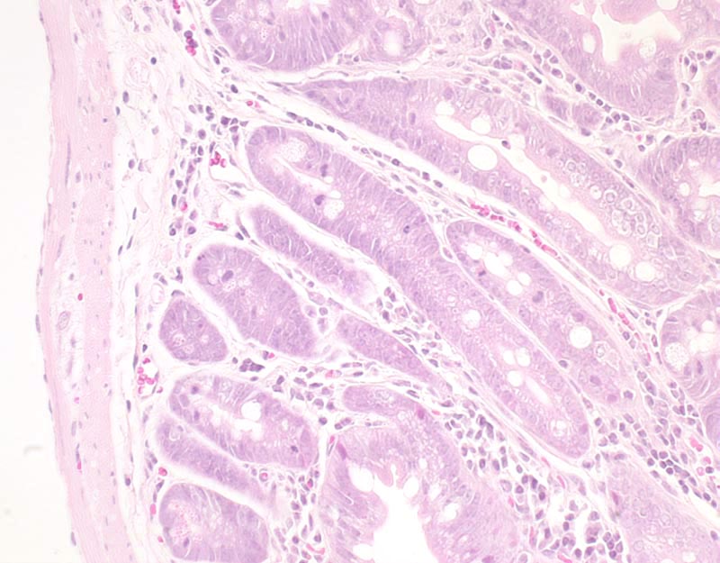
Fig. 3G. Intestinal hamartoma: this is high magnification photomicrograph of the basal portions of an intestinal hamartoma. Cells forming the glands often contain a large apical mucus droplet (goblet cell metaplasia). There is no evidence of atypia or of local invasion. Mitotic rate is high. |
Description |
Hamartoma, polypoid, small intestine |
Age at Necropsy |
280 days |
Contributor |
Miyoshi H (J:76202) |
Pathologist |
Mikaelian I (J:94320) |
Copyright |
Reprinted by permission from the American Association for Cancer Research: Miyoshi H et al., "Gastrointestinal hamartomatous polyposis in Lkb1 heterozygous knockout mice." Cancer Res 2002 Apr 15;62(8):2261-6. |
Method |
H&E |