Pathology Image Detail

Caption

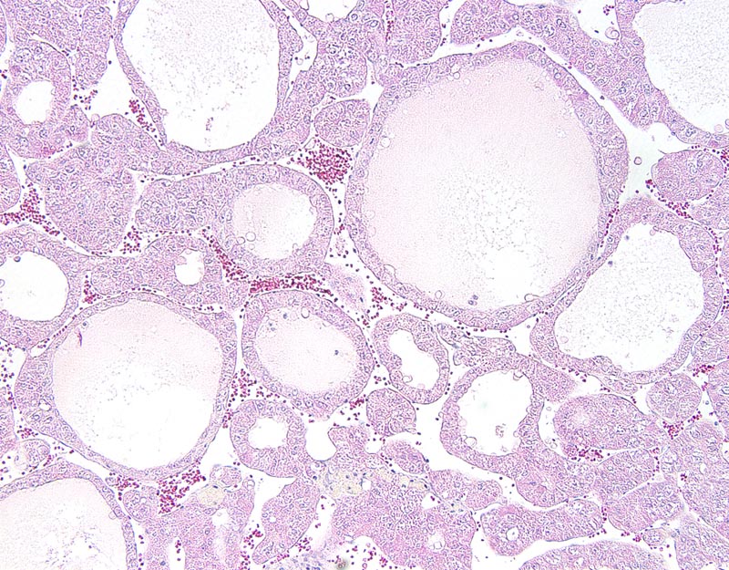
Fig. 1F. Hepatocellular carcinoma, pseudoglandular pattern: the neoplasm is composed of trabeculae with central cavitation and formation of cystic spaces. These cystic spaces generally are filled by a proteinaceous material and they are lined by a one cell-thick cuboidal to low columnar epithelium. Neoplastic cells have distinct cell borders, occasionally apical blebs, and a finely granular acidophilic cytoplasm. The nucleus is central to basal, medium-sized, round to oval, and hypochromatic. Anisokaryosis and anisocytosis are mild.

Description

Carcinoma, pseudoglandular, hepatocellular, liver

Age at Necropsy

unknown

Contributor

Nakau M (J:78502)

Pathologist

Mikaelian I (J:94320)

Copyright

Reprinted by permission from the American Association for Cancer Research: Nakau M et al., "Hepatocellular carcinoma caused by loss of heterozygosity in lkb1 gene knockout mice." Cancer Res 2002 Aug 15;62(16):4549-53.

Method

H&E

Model

MMHC ID
MTB:24829
Tumor Name
Liver • hepatocellular carcinoma - pseudoglandular
Treatment Type
None (spontaneous)
Tumor Synonyms
carcinoma, pseudoglandular, hepatocellular, liver
Organ Affected
Liver
Frequency
observed
Reference
J:78502

Strain

Strain
B6;129-Stk11tm1Mmt/+
Strain Types
targeted mutation (knockout)
General Note
Heterozygous mice. The genetic background was a mix of C57BL/6, 129X1/SvJ, and possibly some other unspecified "129/Sv" substrain.
Strain Synonyms
Lkb1 heterozygote
Lkb1 (+/-)
Lkb1 +/-
Strain Sex
Unspecified
Reproductive Status
reproductive status not specified