Caption |
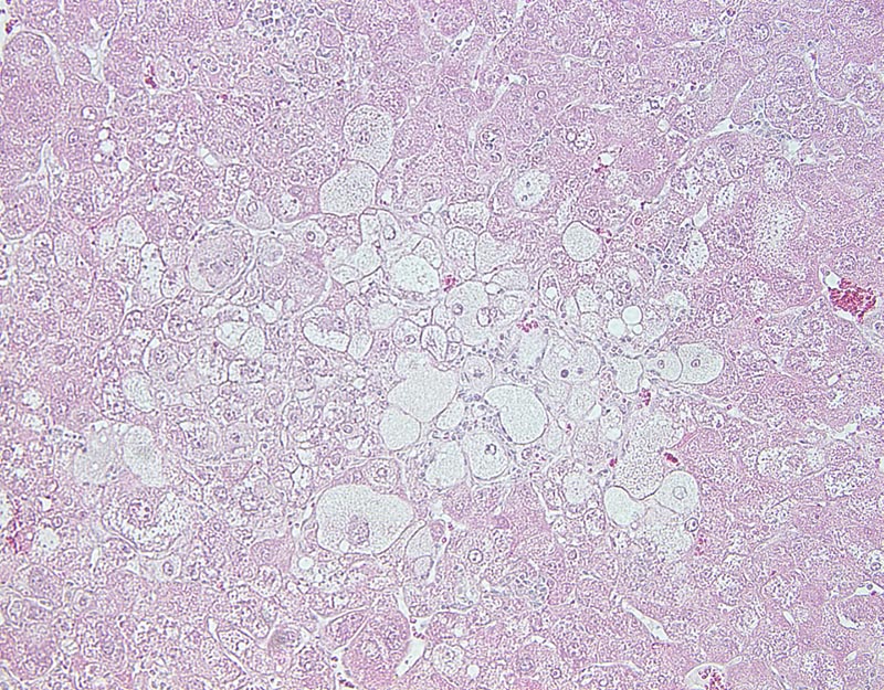
Fig. 1E. Liver, hepatocellular carcinoma: this high magnification photomicrograph shows a neoplasm composed of <2 cells-thick trabeculae and solid areas separated by inconspicuous sinusoids. Neoplastic cells are large to very large, polygonal, with distinct cell borders and a large to very large amount of strongly acidophilic and granular to pale acidophilic and markedly vacuolated cytoplasm. The nucleus is central, oval, medium-sized to large, normochromatic, occasionally with a clumped chromatin and with 1-3 small basophilic nucleoli. Anisokaryosis and anisocytosis are prominent. |