Pathology Image Detail
Caption |
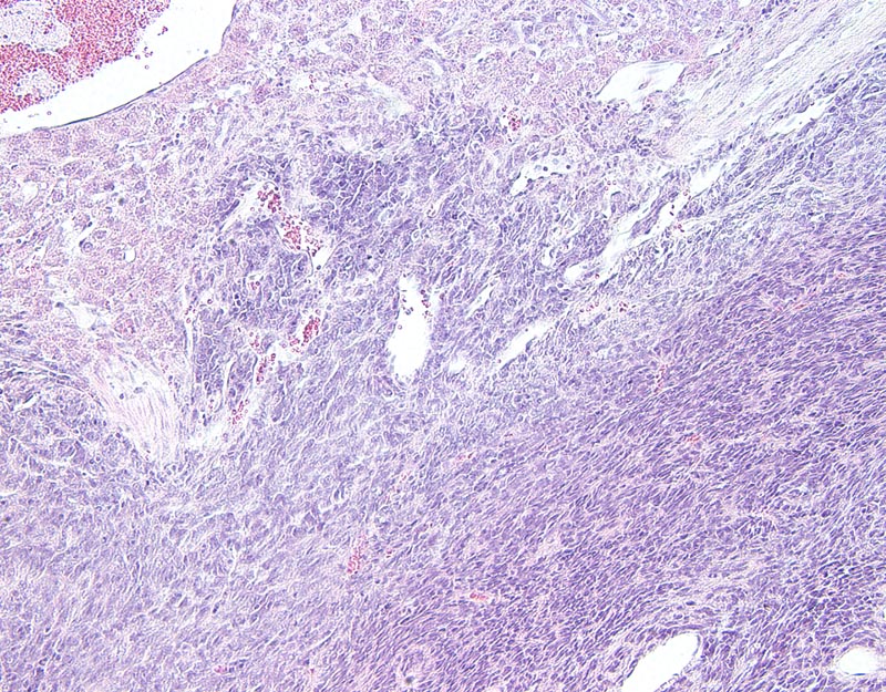
Fig. 1G. The neoplasm is composed of bundles, streams and individual cells supported by a scant amount of fibrovascular stroma. Neoplastic cells are small to medium-sized, with ill-defined to indistinct cell borders, polygonal to spindloid, and with a small amount of basophilic cytoplasm. The nucleus is central, oval to elongated. Anisocytosis is moderate. The neoplasm invades the adjacent hepatic parenchyma (left upper portions of the photomicrograph). |
Description |
Hepatoblastoma, liver |
Age at Necropsy |
unknown |
Contributor |
Nakau M (J:78502) |
Pathologist |
Mikaelian I (J:94320) |
Copyright |
Reprinted by permission from the American Association for Cancer Research: Nakau M et al., "Hepatocellular carcinoma caused by loss of heterozygosity in lkb1 gene knockout mice." Cancer Res 2002 Aug 15;62(16):4549-53. |
Method |
H&E |
Model |
|
Strain |
|