|
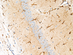
Image Caption:Brain, Hipocampus - Normal tissue. There is strong labeling of glial cells for glial fibrillary acidic protein (GFAP). Glial cells have a dendritic shape. There is moderate non-specific background labeling. The tissues were fixed in Fekete's solution.
|
|
|
Image ID:289 |
|
Source of Image:Mikaelian I |
|
Pathologist:Mikaelian I |
|
Method / Stain:IHC for GFAP |
|
|
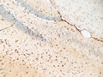
Image Caption:Brain, Hipocampus - Normal tissue. There is strong labeling of glial cells for glial fibrillary acidic protein (GFAP). There is moderate non-specific background labeling. The tissues were fixed in Fekete's solution.
|
|
|
Image ID:288 |
|
Source of Image:Mikaelian I |
|
Pathologist:Mikaelian I |
|
Method / Stain:IHC for GFAP |
|
|
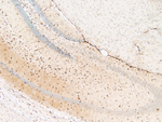
Image Caption:Brain, hipocampus - Normal tissue. There is strong labeling of glial cells for glial fibrillary acidic protein. There is a mild to moderate interstitial background. Antibodies to GFAP are commonly associated with non-specific labeling in epidermal structures. The tissues were fixed in Fekete's solution.
|
|
|
Image ID:287 |
|
Source of Image:Mikaelian I |
|
Pathologist:Mikaelian I |
|
Method / Stain:IHC for GFAP |
|