|
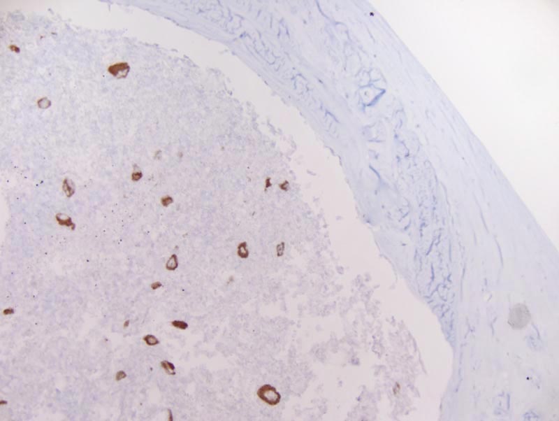
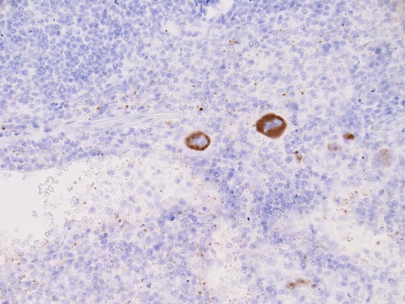
Image Caption:Spleen - normal tissue. There is strong labeling of the cytoplasm of megakaryocytes and of platelets for P-selectin (antibody diluted at 1:25, tissue fixed in IHC Zinc fixative (BD Biosciences), heat-mediated antigen retrieval in 0.1 mM EDTA at pH=7.5). Platelets appear as minute brown dots located in the sinuses of the red pulp and in blood vessels.
|
|
|
Image ID:784 |
|
Source of Image:Mikaelian I |
|
Pathologist:Mikaelian I |
|
Method / Stain:IHC for P-selectin |
|
|
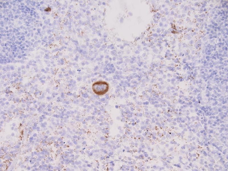
Image Caption:Spleen - normal tissue. There is strong labeling of the cytoplasm of megakaryocytes and of platelets for P-selectin (antibody diluted at 1:50, tissue fixed in 4% paraformaldehyde, heat-mediated antigen retrieval in citrate buffer at pH=6.0). Platelets appear as minute brown dots located in the sinuses of the red pulp and in blood vessels.
|
|
|
Image ID:783 |
|
Source of Image:Mikaelian I |
|
Pathologist:Mikaelian I |
|
Method / Stain:IHC for P-selectin |
|
|
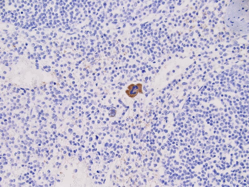
Image Caption:Spleen - normal tissue. There is strong labeling of the cytoplasm of megakaryocytes for P-selectin (antibody diluted at 1:25, tissue fixed in 4% paraformaldehyde, heat-mediated antigen retrieval in 0.1 mM EDTA at pH=7.5). There is weak labeling of the cytoplasm of platelets that appear as minute brown dots located in the sinuses of the red pulp and in blood vessels.
|
|
|
Image ID:782 |
|
Source of Image:Mikaelian I |
|
Pathologist:Mikaelian I |
|
Method / Stain:IHC for P-selectin |
|
|
Image Caption:Spleen - normal tissue. There is strong labeling of the cytoplasm of megakaryocytes and of platelets for P-selectin (antibody diluted at 1:25, tissue fixed in Fekete's solution, heat-mediated antigen retrieval in 0.1 mM EDTA at pH=7.5). Platelets appear as minute brown dots located in the sinuses of the red pulp and in blood vessels.
|
|
|
Image ID:781 |
|
Source of Image:Mikaelian I |
|
Pathologist:Mikaelian I |
|
Method / Stain:IHC for P-selectin |
|
|
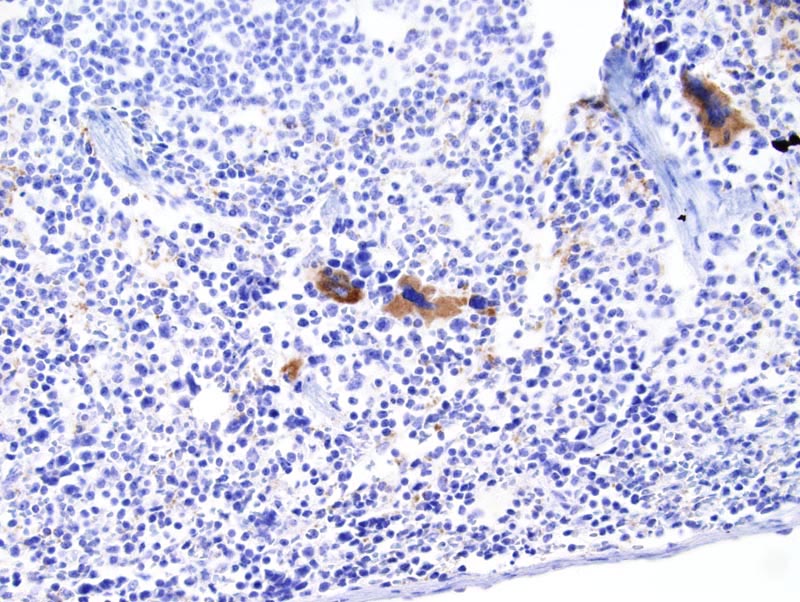
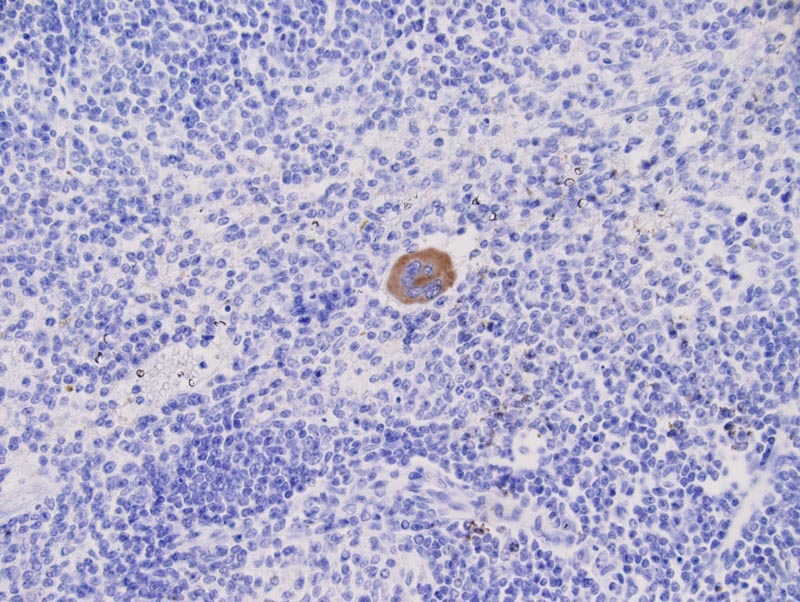
Image Caption:Spleen - normal tissue. There is strong labeling of the cytoplasm of a megakaryocyte for P-selectin (antibody diluted at 1:25, tissue fixed in Fekete's solution, heat-mediated antigen retrieval in citrate buffer at pH=6.0). Platelets are not labeled (platelets, when labeled, appear as minute brown dots located in the sinuses of the red pulp and in blood vessels). Platelets are labeled when the antigen retrieval solution is EDTA instead of citrate buffer.
|
|
|
Image ID:780 |
|
Source of Image:Mikaelian I |
|
Pathologist:Mikaelian I |
|
Method / Stain:IHC for P-selectin |
|