|
| MTB ID |
Tumor Name |
Organ(s) Affected |
Treatment Type |
Agents |
Strain Name |
Strain Sex |
Reproductive Status |
Tumor Frequency |
Age at Necropsy |
Description |
Reference |
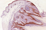
| MTB:17123 |
Eye - Eyelid squamous cell carcinoma |
Eye - Eyelid |
None (spontaneous) |
|
|
Male |
reproductive status not specified |
observed |
510 days |
Eyelid, squamous cell carcinoma |
J:94310 |
|
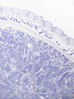
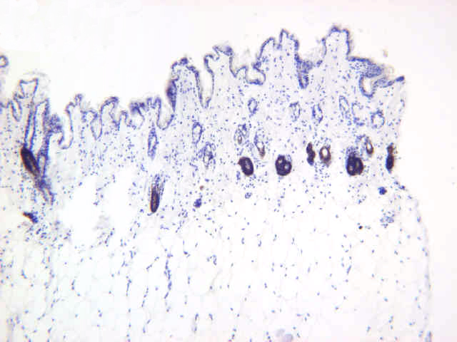
Image Caption:Squamous cell carcinoma of the eyelid. The inner layer of the outer root sheath of normal hair follicles, and not the normal epidermis, express keratin 6. In this neoplasm the suprabasal cells express keratin 6.
|
|
|
Image ID:161 |
|
Source of Image:Mikaelian I |
|
Pathologist:Mikaelian I |
|
Method / Stain:IHC for keratin 6 |
|
|
|
| MTB ID |
Tumor Name |
Organ(s) Affected |
Treatment Type |
Agents |
Strain Name |
Strain Sex |
Reproductive Status |
Tumor Frequency |
Age at Necropsy |
Description |
Reference |
| MTB:29395 |
Mammary gland tumor - type P |
Mammary gland |
None (spontaneous) |
|
|
Female |
reproductive status not specified |
observed |
128 days |
Type P tumor, mammary gland |
J:94320 |
|
Image Caption:Type P tumor: a few neoplastic cells label strongly for keratin 6 in a focus of squamous metaplasia.
|
|
|
Image ID:2085 |
|
Source of Image:Churchill GA |
|
Pathologist:Mikaelian I |
|
Method / Stain:IHC for keratin 6 |
|
|
|
| MTB ID |
Tumor Name |
Organ(s) Affected |
Treatment Type |
Agents |
Strain Name |
Strain Sex |
Reproductive Status |
Tumor Frequency |
Age at Necropsy |
Description |
Reference |
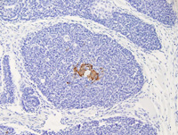
| MTB:29483 |
Mammary gland adenocarcinoma - type P |
Mammary gland |
None (spontaneous) |
|
|
Female |
reproductive status not specified |
observed |
73 days |
Type P tumor, mammary gland |
J:94320 |
|
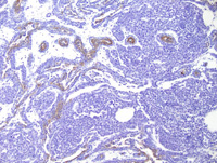
Image Caption:Type P tumor: immunolabelling for keratin 6 is restricted to the suprabasal cells in the areas of ductal differentiation (towards the center of the neoplasm). The fronds of neoplastic cells which mimic terminal end buds of the developing mammary gland do not label for keratin 6.
|
|
|
Image ID:2465 |
|
Source of Image:Churchill GA |
|
Pathologist:Mikaelian I |
|
Method / Stain:IHC for keratin 6 |
|
|
Image Caption:Type P tumor: immunolabelling for keratin 6 is restricted to the suprabasal cells in the areas of ductal differentiation. The fronds of neoplastic cells which mimic terminal end buds of the developing mammary gland do not label for keratin 6.
|
|
|
Image ID:2466 |
|
Source of Image:Churchill GA |
|
Pathologist:Mikaelian I |
|
Method / Stain:IHC for keratin 6 |
|
|
|
| MTB ID |
Tumor Name |
Organ(s) Affected |
Treatment Type |
Agents |
Strain Name |
Strain Sex |
Reproductive Status |
Tumor Frequency |
Age at Necropsy |
Description |
Reference |
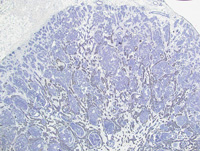
| MTB:29486 |
Mammary gland adenocarcinoma - type P |
Mammary gland |
None (spontaneous) |
|
|
Female |
reproductive status not specified |
observed |
422 days |
Carcinoma, tubular, solid (type P tumor), mammary gland |
J:94320 |
|
Image Caption:Mammary gland: the neoplasm is composed of radiating and branching ducts terminated by club-shaped masses of cells which caricature the terminal end bud of the developing mammary gland. Labeling for keratin 6 is restricted to suprabasal cells in the areas of ductal differentiation.
|
|
|
Image ID:2470 |
|
Source of Image:Churchill GA |
|
Pathologist:Mikaelian I |
|
Method / Stain:IHC for keratin 6 |
|
|
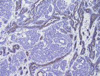
Image Caption:Mammary gland: labeling for keratin 6 is restricted to suprabasal cells in the areas of ductal differentiation.
|
|
|
Image ID:2471 |
|
Source of Image:Churchill GA |
|
Pathologist:Mikaelian I |
|
Method / Stain:IHC for keratin 6 |
|
|
|
| MTB ID |
Tumor Name |
Organ(s) Affected |
Treatment Type |
Agents |
Strain Name |
Strain Sex |
Reproductive Status |
Tumor Frequency |
Age at Necropsy |
Description |
Reference |
| MTB:27738 |
Skin normal tissue (control) |
Skin |
None (spontaneous) |
|
|
Unspecified |
reproductive status not specified |
not applicable |
unknown |
Haired skin - Normal tissue. Keratin 6 immunohistochemistry |
J:94320 |
|
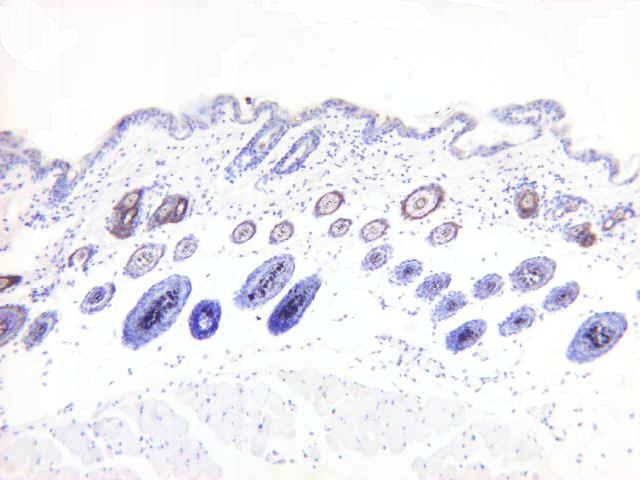
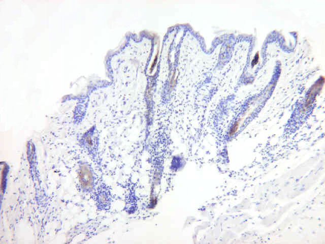
Image Caption:Haired skin - normal tissue. This figure illustrates immunolabeling for keratin 6 (1:6,000 dilution) in a section of the epidermis fixed in 4% paraformaldehyde. There is moderate labeling of the outer root sheath.
|
|
|
Image ID:634 |
|
Source of Image:Mikaelian I |
|
Pathologist:Mikaelian I |
|
Method / Stain:IHC for keratin 6 |
|
|
Image Caption:Haired skin - normal tissue. This figure illustrates immunolabeling for keratin 6 (1:6,000 dilution) in a section of the epidermis fixed in Fekete's solution. There is strong labeling of the outer root sheath.
|
|
|
Image ID:632 |
|
Source of Image:Mikaelian I |
|
Pathologist:Mikaelian I |
|
Method / Stain:IHC for keratin 6 |
|
|
Image Caption:Haired skin - normal tissue. This figure illustrates immunolabeling for keratin 6 (1:6,000 dilution) in a section of the epidermis fixed in Zinc IHC fixative (TM). There is strong labeling of the outer root sheath.
|
|
|
Image ID:633 |
|
Source of Image:Mikaelian I |
|
Pathologist:Mikaelian I |
|
Method / Stain:IHC for keratin 6 |
|
|
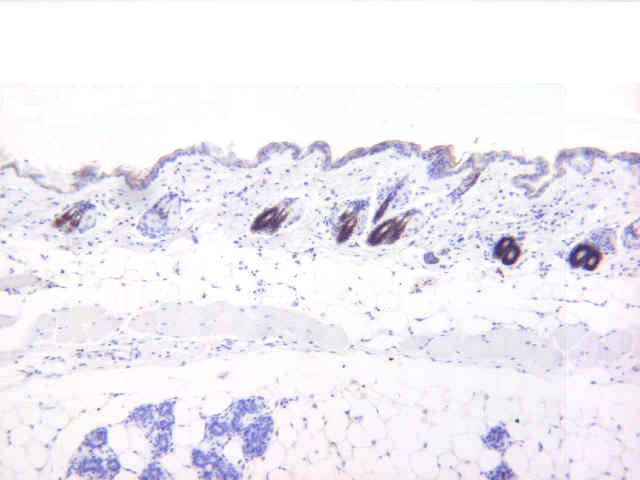
Image Caption:Haired skin - normal tissue. This figure illustrates immunolabeling for keratin 6 (1:6,000 dilution) in a section of the epidermis fixed in 10% neutral buffered formalin. There is mild to moderate labeling of the outer root sheath. There is also moderate lymphocytic mural folliculitis, which is diagnosis for alopecia areata.
|
|
|
Image ID:635 |
|
Source of Image:Mikaelian I |
|
Pathologist:Mikaelian I |
|
Method / Stain:IHC for keratin 6 |
|
|