|
| MTB ID |
Tumor Name |
Organ(s) Affected |
Treatment Type |
Agents |
Strain Name |
Strain Sex |
Reproductive Status |
Tumor Frequency |
Age at Necropsy |
Description |
Reference |
| MTB:27693 |
Skin normal tissue (control) |
Skin |
None (spontaneous) |
|
|
Unspecified |
reproductive status not specified |
not applicable |
35 days |
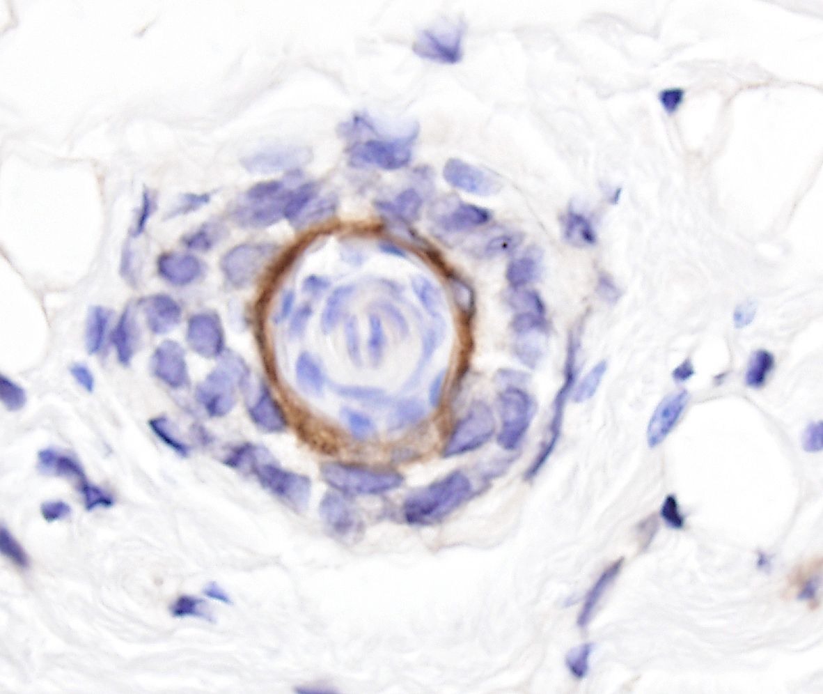
Haired skin - Normal tissue. Keratins 5 and 8 immunohistochemistry, non-specific labeling |
J:94320 |
Image Caption:Haired skin - normal tissue. A cross section of a hair follicle shows strong labeling of the inner layer of the outer root sheath for keratins 5/8 (antibody diluted at 1:100, tissue fixed in Fekete's solution, enzymatic digestion in trypsin). Keratin 5 is a marker of basal cells while keratin 8 is a marker of simple epithelia. Hence labeling in this case is interpreted as the result of a cross-reaction of the antibody with undetermined proteins.
NOTE: Haired skin, esophageal mucosa, and oral mucosa can be used as positive control for this antibody. However, this antibody does not label all the structures it is supposed to label based on reported expression patterns for keratins 5 and 8.
|
|
|
Image ID:642 |
|
Source of Image:Mikaelian I |
|
Pathologist:Mikaelian I |
|
Method / Stain:IHC for keratin 5 and 8 |
|
|
|
| MTB ID |
Tumor Name |
Organ(s) Affected |
Treatment Type |
Agents |
Strain Name |
Strain Sex |
Reproductive Status |
Tumor Frequency |
Age at Necropsy |
Description |
Reference |
| MTB:27707 |
Tongue normal tissue (control) |
Tongue |
None (spontaneous) |
|
|
Unspecified |
reproductive status not specified |
not applicable |
35 days |
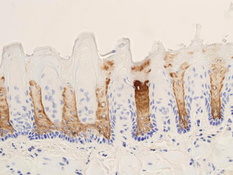
Tongue - Normal tissue. Keratins 5 and 8 immunohistochemistry, non-specific labeling |
J:94320 |
|
Image Caption:Oral mucosa (tongue) - normal tissue. There is strong labeling for keratins 5/8 of suprabasal cells of the inter-papillary mucosa (antibody diluted at 1:500, tissue fixed in 4% paraformaldehyde, enzymatic digestion in trypsin). Keratin 5 is a marker of basal cells while keratin 8 is a marker of simple epithelia. Hence labeling in this case is interpreted as the result of a cross-reaction of the antibody with undetermined proteins.
|
|
|
Image ID:1352 |
|
Source of Image:Mikaelian I |
|
Pathologist:Mikaelian I |
|
Method / Stain:IHC for keratin 5 and 8 |
|
|
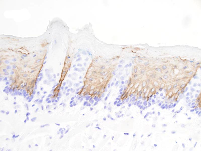
Image Caption:Oral mucosa (tongue) - normal tissue. There is strong labeling for keratins 5/8 of suprabasal cells of the inter-papillary mucosa (antibody diluted at 1:500, tissue fixed in Fekete's solution, enzymatic digestion in trypsin). Keratin 5 is a marker of basal cells while keratin 8 is a marker of simple epithelia. Hence labeling in this case is interpreted as the result of a cross-reaction of the antibody with undetermined proteins.
|
|
|
Image ID:1351 |
|
Source of Image:Mikaelian I |
|
Pathologist:Mikaelian I |
|
Method / Stain:IHC for keratin 5 and 8 |
|
|