|
| MTB ID |
Tumor Name |
Organ(s) Affected |
Treatment Type |
Agents |
Strain Name |
Strain Sex |
Reproductive Status |
Tumor Frequency |
Age at Necropsy |
Description |
Reference |
| MTB:27648 |
Intestine normal tissue (control) |
Intestine |
None (spontaneous) |
|
|
Unspecified |
reproductive status not specified |
not applicable |
unknown |
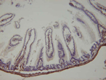
Intestine - Smooth muscle - normal tissue. |
J:94310 |
|
Image Caption:Colon - Normal tissue. There is intense cytoplasmic labelling for alpha smooth muscle actin of smooth muscle fibers of the muscularis mucosae and of the wall of the colon (antibody diluted at 1:6,000, tissue fixed in Bouin's solution).
|
|
|
Image ID:1102 |
|
Source of Image:Mikaelian I |
|
Pathologist:Mikaelian I |
|
Method / Stain:IHC for alpha smooth muscle actin |
|
|
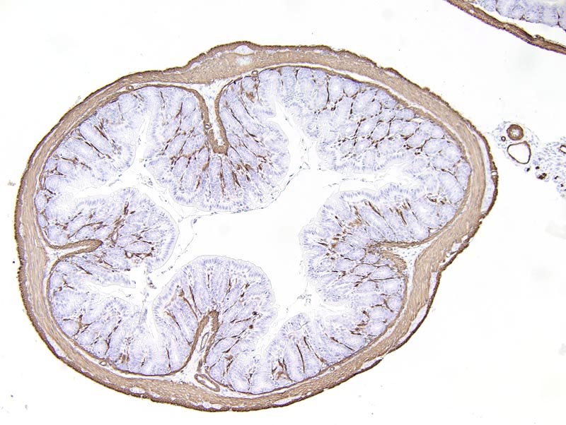
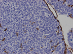
Image Caption:Intestine - Smooth muscle - Normal tissue. There is intense labelling of smooth muscle fibers located in intestinal villi and in the wall of the intestine. There is mild interstitial background labelling. The tissue was fixed in Fekete's solution and labeled with an antibody diluted at 1:40,000.
|

|
|
Image ID:231 |
|
Source of Image:Mikaelian I |
|
Pathologist:Mikaelian I |
|
Method / Stain:IHC for alpha smooth muscle actin |
|
|
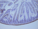
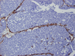
Image Caption:Intestine - Smooth muscle - Normal tissue. There is strong labeling of smooth muscle fibers located in intestinal villi and in the wall of the intestine. There is no interstitial background labelling. The tissue was fixed in Bouin's fixative and labeled with an antibody diluted at 1:40,000.
|
|
|
Image ID:233 |
|
Source of Image:Mikaelian I |
|
Pathologist:Mikaelian I |
|
Method / Stain:IHC for alpha smooth muscle actin |
|
|
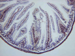
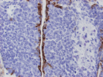
Image Caption:Intestine - Smooth muscle - Normal tissue. There is intense labeling of smooth muscle fibers located in intestinal villi and in the wall of the intestine. There is no background labeling. The tissue was fixed in Zinc buffer and labelled with an antibody diluted at 1:40,000.
|
|
|
Image ID:235 |
|
Source of Image:Mikaelian I |
|
Pathologist:Mikaelian I |
|
Method / Stain:IHC for alpha smooth muscle actin |
|
|
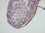
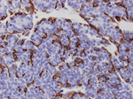
Image Caption:Intestine - Smooth muscle - Normal tissue. There is strong labeling of smooth muscle fibers located in intestinal villi and in the wall of the intestine. There is moderate interstitial background labeling. The tissue was fixed in 10% neutral buffered formalin and labeled with an antibody diluted at 1:40,000.
|
|
|
Image ID:237 |
|
Source of Image:Mikaelian I |
|
Pathologist:Mikaelian I |
|
Method / Stain:IHC for alpha smooth muscle actin |
|
|
Image Caption:Intestine - Smooth muscle - Normal tissue. There is strong labeling of smooth muscle fibers located in intestinal villi and in the wall of the intestine. There is mild interstitial background labeling. The tissue was fixed in 4% paraformaldehyde and labeled with an antibody diluted at 1:40,000.
|
|
|
Image ID:239 |
|
Source of Image:Mikaelian I |
|
Pathologist:Mikaelian I |
|
Method / Stain:IHC for alpha smooth muscle actin |
|
|
|
| MTB ID |
Tumor Name |
Organ(s) Affected |
Treatment Type |
Agents |
Strain Name |
Strain Sex |
Reproductive Status |
Tumor Frequency |
Age at Necropsy |
Description |
Reference |
| MTB:18035 |
Mammary gland adenocarcinoma |
Mammary gland |
None (spontaneous) |
|
|
Female |
reproductive status not specified |
observed |
210 days |
Mammary gland, adenocarcinoma. |
J:94310 |
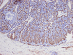
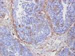
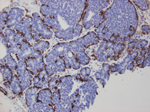
Image Caption:Mammary gland, adenocarcinoma.
Cells located at the periphery of neoplastic trabeculae and lobules show intense immunolabeling for alpha smooth muscle actin, which indicates myoepithelial differentiation.
|
|
|
Image ID:209 |
|
Source of Image:Mikaelian I |
|
Pathologist:Mikaelian I |
|
Method / Stain:IHC for alpha smooth muscle actin |
|
|
|
| MTB ID |
Tumor Name |
Organ(s) Affected |
Treatment Type |
Agents |
Strain Name |
Strain Sex |
Reproductive Status |
Tumor Frequency |
Age at Necropsy |
Description |
Reference |
| MTB:18036 |
Mammary gland adenocarcinoma |
Mammary gland |
None (spontaneous) |
|
|
Female |
reproductive status not specified |
observed |
186 days |
Mammary gland, adenocarcinoma. |
J:94310 |
|
Image Caption:Mammary gland, adenocarcinoma. A continuous row of neoplastic cells, located at the periphery of neoplastic lobules, labels for alpha smooth muscle actin.
|
|
|
Image ID:222 |
|
Source of Image:Mikaelian I |
|
Pathologist:Mikaelian I |
|
Method / Stain:IHC for alpha smooth muscle actin |
|
|
Image Caption:Mammary gland, adenocarcinoma. Smooth muscle fibers of blood vessels within the neoplasm label intensely for alpha smooth muscle actin.
|
|
|
Image ID:220 |
|
Source of Image:Mikaelian I |
|
Pathologist:Mikaelian I |
|
Method / Stain:IHC for alpha smooth muscle actin |
|
|
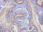
Image Caption:Mammary gland, adenocarcinoma. Neoplastic cells labeling for alpha smooth muscle actin form a continuous row at the periphery of neoplastic lobules in this area with a comedo pattern. In addition, a moderate number of neoplastic cells randomly distributed in neoplastic lobules moderately label for alpha smooth muscle actin. A moderate number of myofibroblasts scattered in the stroma of the tumor and smooth muscle fibers of the wall of blood vessels also label for alpha smooth muscle actin.
|
|
|
Image ID:221 |
|
Source of Image:Mikaelian I |
|
Pathologist:Mikaelian I |
|
Method / Stain:IHC for alpha smooth muscle actin |
|
|
|
| MTB ID |
Tumor Name |
Organ(s) Affected |
Treatment Type |
Agents |
Strain Name |
Strain Sex |
Reproductive Status |
Tumor Frequency |
Age at Necropsy |
Description |
Reference |
| MTB:18037 |
Mammary gland adenocarcinoma |
Lung |
None (spontaneous) |
|
|
Female |
reproductive status not specified |
observed |
186 days |
Lung: Mammary adenocarcinoma, metastatic to the lung. |
J:94310 |
|
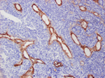
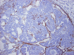
Image Caption:Lung: mammary adenocarcinoma, metastatic to the lung. This figure represents a low magnification image of a mammary adenocarcinoma metastatic to the lung and immunolabeled for alpha smooth muscle actin. A few scattered neoplastic cells and smooth muscle fibers of the wall of blood vessels of the stroma strongly label for smooth muscle actin. Labeling of neoplastic cells indicates myoepithelial differentiation.
|
|
|
Image ID:211 |
|
Source of Image:Mikaelian I |
|
Pathologist:Mikaelian I |
|
Method / Stain:IHC for alpha smooth muscle actin |
|
|
Image Caption:Mammary adenocarcinoma, metastatic to the lung, tubular area. Labeling for alpha smooth muscle actin is restricted to basal cells in this area of tubular differentiation. This feature indicates myoepithelial differentiation.
|
|
|
Image ID:212 |
|
Source of Image:Mikaelian I |
|
Pathologist:Mikaelian I |
|
Method / Stain:IHC for alpha smooth muscle actin |
|
|
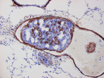
Image Caption:Mammary adenocarcinoma, metastatic to the lung. A neoplastic embolus originating from a mammary tumor is present in an ectatic pulmonary artery. Scattered neoplastic cells within the embolus intensely label for alpha smooth muscle actin, which indicates myoepithelial differentiation. There is also intense labeling of smooth muscle fibers of the wall of the artery.
|
|
|
Image ID:210 |
|
Source of Image:Mikaelian I |
|
Pathologist:Mikaelian I |
|
Method / Stain:IHC for alpha smooth muscle actin |
|
|
|
| MTB ID |
Tumor Name |
Organ(s) Affected |
Treatment Type |
Agents |
Strain Name |
Strain Sex |
Reproductive Status |
Tumor Frequency |
Age at Necropsy |
Description |
Reference |
| MTB:18038 |
Mammary gland adenocarcinoma |
Mammary gland |
None (spontaneous) |
|
|
Female |
reproductive status not specified |
observed |
173 days |
Mammary gland, adenocarcinoma. |
J:94310 |
|
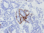
Image Caption:Mammary gland, adenocarcinoma. A continuous row of neoplastic cells at the periphery of a neoplastic lobule label intensely for alpha smooth muscle actin, which indicates myoepithelial differentiation. Scattered neoplastic cells throughout the lobule and smooth muscle fibers in the wall of a small vein also label strongly for alpha smooth muscle actin.
|
|
|
Image ID:217 |
|
Source of Image:Mikaelian I |
|
Pathologist:Mikaelian I |
|
Method / Stain:IHC for alpha smooth muscle actin |
|
|
Image Caption:Mammary gland, adenocarcinoma. Neoplastic cells at the periphery of large lobules in an area with a comedo pattern strongly label for alpha smooth muscle actin. A few neoplastic cells scattered throughout neoplastic lobules show moderate labeling.
|
|
|
Image ID:219 |
|
Source of Image:Mikaelian I |
|
Pathologist:Mikaelian I |
|
Method / Stain:IHC for alpha smooth muscle actin |
|
|
Image Caption:Mammary gland, adenocarcinoma. Neoplastic cells at the periphery of neoplastic lobules intensely label for alpha smooth muscle actin, which indicates myoepithelial differentiation. This picture is taken from an area where the neoplasm had a solid to comedo-type pattern.
|
|
|
Image ID:215 |
|
Source of Image:Mikaelian I |
|
Pathologist:Mikaelian I |
|
Method / Stain:IHC for alpha smooth muscle actin |
|
|
Image Caption:Mammary gland, adenocarcinoma. Neoplastic cells located at the periphery of tubular structures intensely label for smooth muscle actin in this area with a tubular pattern. Alpha smooth muscle actin expression is an indication of smooth muscle differentiation.
|
|
|
Image ID:216 |
|
Source of Image:Mikaelian I |
|
Pathologist:Mikaelian I |
|
Method / Stain:IHC for alpha smooth muscle actin |
|
|
Image Caption:Mammary gland, adenocarcinoma. This figure represent an area of transition between an acinar pattern (lower left) and a solid pattern (upper right). Neoplastic cells at the periphery of the acini and thick trabeculae label strongly for alpha smooth muscle actin, which indicates myoepithelial differentiation.
|
|
|
Image ID:218 |
|
Source of Image:Mikaelian I |
|
Pathologist:Mikaelian I |
|
Method / Stain:IHC for alpha smooth muscle actin |
|
|