|
| MTB ID |
Tumor Name |
Organ(s) Affected |
Treatment Type |
Agents |
Strain Name |
Strain Sex |
Reproductive Status |
Tumor Frequency |
Age at Necropsy |
Description |
Reference |
| MTB:27719 |
Esophagus normal tissue (control) |
Esophagus |
None (spontaneous) |
|
|
Unspecified |
reproductive status not specified |
not applicable |
unknown |
Esophagus - Normal tissue. p63 immunohistochemistry |
J:94320 |
|
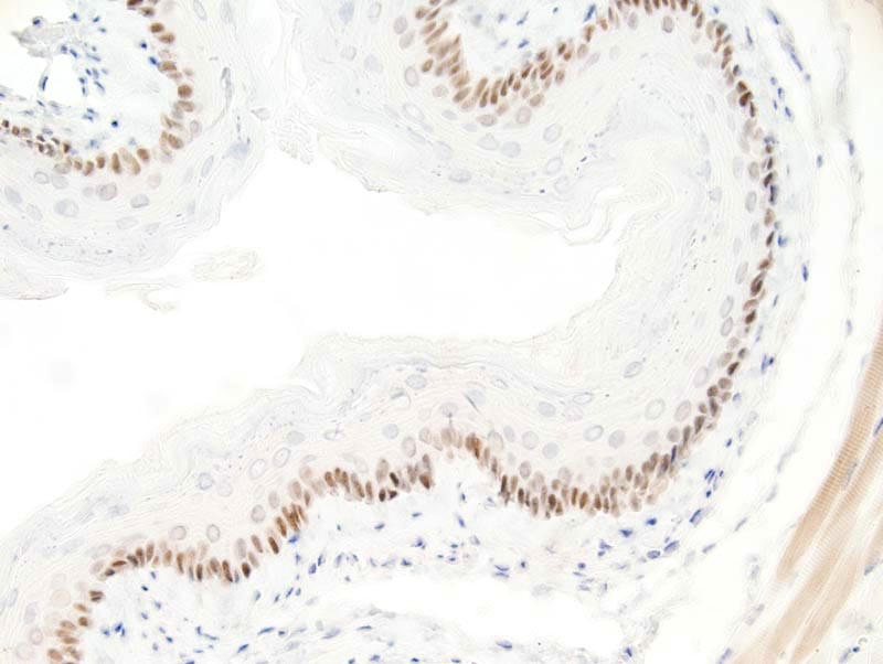
Image Caption:Esophagus - normal tissue. There is strong nuclear labeling for p63 of basal cells of the esophagus (antibody diluted at 1:25, tissue fixed in Zinc IHC fixative, heat-mediated antigen retrieval in 0.1mM EDTA at pH=7.5).There is moderate artifactual labeling of the cytoplasm of skeletal muscle fibers located at the vicinity of the esophaus.
|
|
|
Image ID:593 |
|
Source of Image:Mikaelian I |
|
Pathologist:Mikaelian I |
|
Method / Stain:IHC for p63 |
|
|
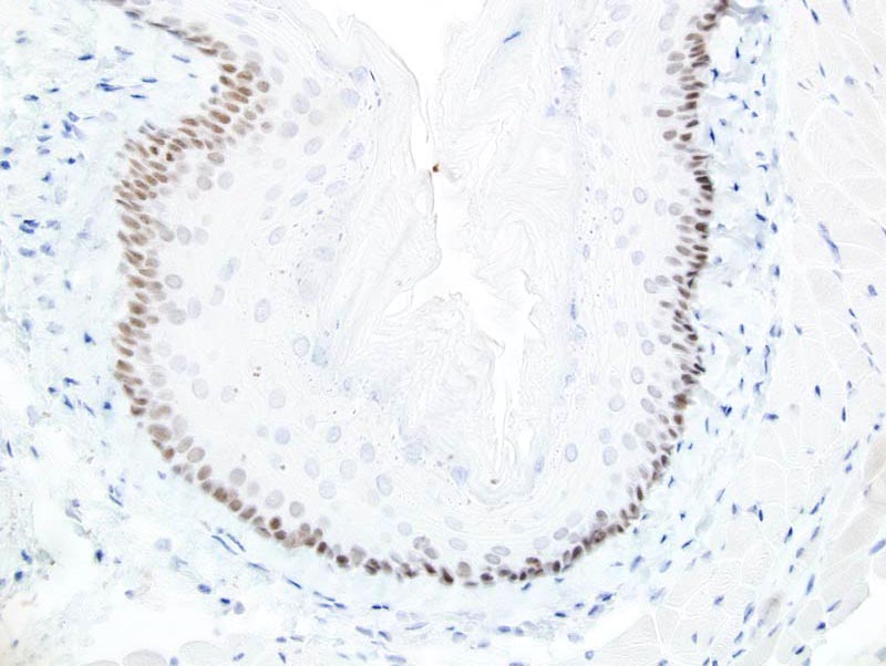
Image Caption:Esophagus - normal tissue. There is strong nuclear labeling for p63 of basal cells of the esophagus (antibody diluted at 1:25, tissue fixed in 10% neutral buffered formalin, heat-mediated antigen retrieval in 0.1mM EDTA at pH=7.5).
|
|
|
Image ID:592 |
|
Source of Image:Mikaelian I |
|
Pathologist:Mikaelian I |
|
Method / Stain:IHC for p63 |
|
|
|
| MTB ID |
Tumor Name |
Organ(s) Affected |
Treatment Type |
Agents |
Strain Name |
Strain Sex |
Reproductive Status |
Tumor Frequency |
Age at Necropsy |
Description |
Reference |
| MTB:46353 |
Prostate gland prostatic intraepithelial neoplasia - high grade (HGPIN) |
Prostate gland |
None (spontaneous) |
|
|
Male |
reproductive status not specified |
84% |
16 months |
immunohistochemistry various markers in PIN from Brca2;Trp53 homozygous mutant mice compared to normal control prostate |
J:161511 |
|
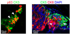
Image Caption:Fig. 4D&E "Left panel shows p63 (red) and CK5 (green) fluorescent immunohistochemistry analysis with labelled cells protruding into the lumen (white arrowheads) in a region of PIN in Brca2F/F;Trp53F/F;PBCre4 mutants. Right panel shows CK5 (green, basal cells) and CK8 (red, luminal cells) fluorescent immunohistochemistry with PIN lesions in Brca2F/F;Trp53F/F;PBCre4 mutants displaying an increase in luminal cells next to clusters of basal cells. White arrowhead marks CK5 and CK8 double-labelled cells. DAPI nuclear stain is blue. The anterior prostates of 16-month-old animals are shown."
|
|
|
Image ID:4582 |
|
Source of Image:Francis JC |
|
Pathologist:Francis JC |
|
Method / Stain:IHC for various markers |
|
|
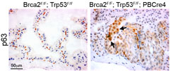
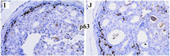
Image Caption:Fig. 4C "p63 immunohistochemistry shows an increase in p63-expressing cells in HG PIN lesions in Brca2F/F;Trp53F/F;PBCre4 mutant prostates and normal expression in the basal cells of control (Brca2F/F;Trp53F/F) prostates. Arrows indicate a cluster of abnormal p63-expressing cells that are rounder and nearer the lumen. ... The anterior prostates of 16-month-old animals are shown."
|
|
|
Image ID:4581 |
|
Source of Image:Francis JC |
|
Pathologist:Francis JC |
|
Method / Stain:IHC for Trp63 |
|
|
|
| MTB ID |
Tumor Name |
Organ(s) Affected |
Treatment Type |
Agents |
Strain Name |
Strain Sex |
Reproductive Status |
Tumor Frequency |
Age at Necropsy |
Description |
Reference |
| MTB:60992 |
Prostate gland prostatic intraepithelial neoplasia - high grade (HGPIN) |
Prostate gland |
None (spontaneous) |
|
|
Male |
reproductive status not specified |
100% |
|
high-grade PIN; H&E and immunohistochemical analysis |
J:195810 |
|
Image Caption:Fig. 5I,J Immunohistochemistry for TRP63 "performed on prostate tissues of 4-week-old PtenloxP/loxP:Osr1-Cre mice."
|
|
|
Image ID:5278 |
|
Source of Image:Kwak MK |
|
Pathologist:Kwak MK |
|
Method / Stain:IHC for TRP63 |
|
|
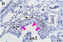
Image Caption:Fig. 5R Immunohistochemistry for TRP63 in prostate tissues of 4-week-old PtenloxP/loxP:Osr1-Cre mice.
|
|
|
Image ID:5283 |
|
Source of Image:Kwak MK |
|
Pathologist:Kwak MK |
|
Method / Stain:IHC for TRP63 |
|
|
|
| MTB ID |
Tumor Name |
Organ(s) Affected |
Treatment Type |
Agents |
Strain Name |
Strain Sex |
Reproductive Status |
Tumor Frequency |
Age at Necropsy |
Description |
Reference |
| MTB:61038 |
Prostate gland prostatic intraepithelial neoplasia - high grade (HGPIN) |
Prostate gland |
None (spontaneous) |
|
|
Male |
reproductive status not specified |
observed |
|
high-grade PIN; immunohistochemical analysis of Pten and Trp63 in adjacent slides |
J:195810 |
|
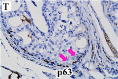
Image Caption:Fig. 5T Immunohistochemistry for TRP63 in prostate tissues of 4-week-old PtenloxP/loxP:PB-Cre4 mice.
|
|
|
Image ID:5285 |
|
Source of Image:Kwak MK |
|
Pathologist:Kwak MK |
|
Method / Stain:IHC for TRP63 |
|
|
|
| MTB ID |
Tumor Name |
Organ(s) Affected |
Treatment Type |
Agents |
Strain Name |
Strain Sex |
Reproductive Status |
Tumor Frequency |
Age at Necropsy |
Description |
Reference |
| MTB:27709 |
Trachea normal tissue (control) |
Trachea |
None (spontaneous) |
|
|
Unspecified |
reproductive status not specified |
not applicable |
35 days |
Trachea - Normal tissue. p63 immunohistochemistry |
J:94320 |
|
Image Caption:Trachea - normal tissue. There is moderate nuclear labeling for p63 of basal cells of the trachea (antibody diluted at 1:25, tissue fixed in 10% neutral buffered formalin, heat-mediated antigen retrieval in 0.1mM EDTA buffer at pH=7.5).
|
|
|
Image ID:918 |
|
Source of Image:Mikaelian I |
|
Pathologist:Mikaelian I |
|
Method / Stain:IHC for p63 |
|
|