|
| MTB ID |
Tumor Name |
Organ(s) Affected |
Treatment Type |
Agents |
Strain Name |
Strain Sex |
Reproductive Status |
Tumor Frequency |
Age at Necropsy |
Description |
Reference |
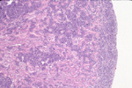
| MTB:18408 |
Blood vessel hemangioma - cavernous |
Skin |
None (spontaneous) |
|
|
Female |
reproductive status not specified |
observed |
155 days |
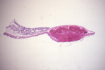
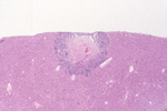
Haired skin, dermal hemangioma, cavernous. |
J:94314 |
|
Image Caption:This is a large, nodular, unencapsulated, well-delineated, poor cellular, ulcerated dermal neoplasm. This neoplasm is made of blood-filed cavernous vascular spaces.
|
|
|
Image ID:385 |
|
Source of Image:Mikaelian I |
|
Pathologist:Mikaelian I |
|
Method / Stain:H&E |
|
|
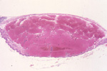
Image Caption:This is a large, nodular, unencapsulated, well-delineated, poorly cellular, ulcerated dermal neoplasm. This neoplasm is made of blood-filled cavernous vascular spaces.
|
|
|
Image ID:386 |
|
Source of Image:Mikaelian I |
|
Pathologist:Mikaelian I |
|
Method / Stain:H&E |
|
|
|
| MTB ID |
Tumor Name |
Organ(s) Affected |
Treatment Type |
Agents |
Strain Name |
Strain Sex |
Reproductive Status |
Tumor Frequency |
Age at Necropsy |
Description |
Reference |
| MTB:18499 |
Bone osteosarcoma |
Bone |
None (spontaneous) |
|
|
Male |
reproductive status not specified |
observed |
147 days |
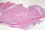
Bone - Femur, osteosarcoma, osteoblastic |
J:94314 |
|
Image Caption:A large, modular, unencapsulated, invasive neoplastic mass arose from the femur and invaded the nearby skeletal muscle.
|
|
|
Image ID:400 |
|
Source of Image:Mikaelian I |
|
Pathologist:Mikaelian I |
|
Method / Stain:H&E |
|
|
|
| MTB ID |
Tumor Name |
Organ(s) Affected |
Treatment Type |
Agents |
Strain Name |
Strain Sex |
Reproductive Status |
Tumor Frequency |
Age at Necropsy |
Description |
Reference |
| MTB:27563 |
Germ cell (sex not specified) teratocarcinoma |
Germ cell (sex not specified) |
None (spontaneous) |
|
|
Unspecified |
reproductive status not specified |
observed |
35 days |
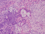
Teratocarcinoma (malignant teratoma), extra-gonadal |
J:94314 |
|
Image Caption:The neoplasm shows epithelial (left portion of the photomicrograph) and neural (right portion of the photomicrograph). An epithelial structure at the center of the photomicrograph is distended by a mucoid substance surrounded by a one cell-thick ciliated cuboidal epithelium reminiscent of the epithelium of the respiratory tract. Vacuolated cells may represent areas of chondromatous or lipomatous differentiation. Scattered areas of skeletal muscle are also present.
|
|
|
Image ID:428 |
|
Source of Image:Wu LC |
|
Pathologist:Mikaelian I |
|
Method / Stain:H&E |
|
|
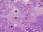
Image Caption:This photomicrograph illustrates areas of osteo-chondromatous (OC), neural (N), epithelial (E), and skeletal muscle (M) differentiation. Areas of osteo-chondromatous differentiation are characterized by the formation of cartilaginous masses with endochondral ossification. Areas of skeletal muscle differentiation are composed of bundles of immature myocytes separated by small amounts of fibrovascular stroma. Areas of epithelial differentiation are composed of trabeculae, nests and acini. Some of the acini are surrounded by goblet cells and are moderately distended by a mucoid substance. Areas of neural differentiation are composed of glial cells and neurones scattered in a pale acidophilic substance consistent with glia.
|
|
|
Image ID:423 |
|
Source of Image:Wu LC |
|
Pathologist:Mikaelian I |
|
Method / Stain:H&E |
|
|
Image Caption:This portion of the neoplasm comprises a large island of chondromatous differentiation. It is surrounded by areas of epithelial differentiation characterized by the formation of solid areas (predominantly), trabeculae, glands and tubules. A few small areas of neural differentiation are interspersed between the areas of chondromatous and epithelial differentiation.
|
|
|
Image ID:426 |
|
Source of Image:Wu LC |
|
Pathologist:Mikaelian I |
|
Method / Stain:H&E |
|
|
Image Caption:This portion of the neoplasm contains an island of cartilage with endochondral ossification (lower right corner), an epithelial structure with squamous differentiation and a prominent stratum granulosum (upper left corner), and an area of tubular differentiation (lower left corner). These structures are separated by areas of skeletal muscle differentiation composed of closely packed streaming bundles of fusiforms cells with eccentric nuclei.
|
|
|
Image ID:424 |
|
Source of Image:Wu LC |
|
Pathologist:Mikaelian I |
|
Method / Stain:H&E |
|
|
Image Caption:This is a nodular, unencapsulated, locally invasive, densely cellular neoplastic mass that arose in the right axillary region. Large areas of liquefaction necrosis (pale acidophilic portions) are scattered throughout the neoplasm.
|
|
|
Image ID:421 |
|
Source of Image:Wu LC |
|
Pathologist:Mikaelian I |
|
Method / Stain:H&E |
|
|
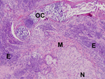
Image Caption:The neoplasm contains areas of osteo-chondromatous (OC), neural (N), skeletal muscle (M) and epithelial (E) differentiation. The areas of osteo-chondromatous differentiation are associated with hematopoiesis. The areas of epithelial differentiation comprise tubular/glandular and solid portions.
|
|
|
Image ID:422 |
|
Source of Image:Wu LC |
|
Pathologist:Mikaelian I |
|
Method / Stain:H&E |
|
|
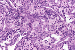
Image Caption:The tumor, in this portion, shows epithelial and neuro-endocrine differentiation. One area of epithelial differentiation is characterized by the formation of a dilated tubule lined by a one cell-thick columnar epithelium reminiscent of the respiratory epithelium. However, most epithelial areas are composed of thick trabeculae and tubules lined by a pluristratified epithelium. These cells are polygonal to columnar and have a moderate amount of basophilic to acidophilic cytoplasm. Neuroendocrine-like areas are characterized by the formation of small nests composed of columnar to fusiform cells with a pale acidophilic cytoplasm. Mitotic index is high and numerous neoplastic cells undergo single cell necrosis.
|
|
|
Image ID:429 |
|
Source of Image:Wu LC |
|
Pathologist:Mikaelian I |
|
Method / Stain:H&E |
|
|
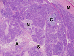
Image Caption:This portion of the neoplasm comprises portions with chondromatous (C), neural (N), adipose (A), and skeletal muscle muscle (S) differentiation separated by cords, trabeculae and nests of epithelial cells which represent ?75% of the tumor. A pre-existing skeletal muscle (M) is pushed aside by neoplasm.
|
|
|
Image ID:425 |
|
Source of Image:Wu LC |
|
Pathologist:Mikaelian I |
|
Method / Stain:H&E |
|
|
Image Caption:A large island of chondromatous differentiation is surrounded by areas of epithelial differentiation. On the right lower corner, neoplastic cells for a dilated tubule lined by a one cell-thick ciliated epithelium reminiscent of the respiratory epithelium.
|
|
|
Image ID:427 |
|
Source of Image:Wu LC |
|
Pathologist:Mikaelian I |
|
Method / Stain:H&E |
|
|
|
| MTB ID |
Tumor Name |
Organ(s) Affected |
Treatment Type |
Agents |
Strain Name |
Strain Sex |
Reproductive Status |
Tumor Frequency |
Age at Necropsy |
Description |
Reference |
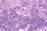
| MTB:18470 |
Leukocyte - Lymphocyte lymphoma |
Kidney |
None (spontaneous) |
|
|
Male |
reproductive status not specified |
observed |
149 days |
Lymphoma. |
J:94314 |
|
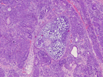
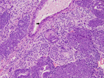
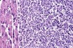
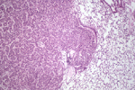
Image Caption:Kidney, lymphoma: The interstitium of the kidney and the subcapsular area are markedly expanded by sheets of closely-packed monotonous round cells.
|
|
|
Image ID:390 |
|
Source of Image:Mikaelian I |
|
Pathologist:Mikaelian I |
|
Method / Stain:H&E |
|
|
Image Caption:Kidney, lymphoma: Neoplastic cells form closely-packed sheets which expend the interstitium. Neoplastic cells are round, with indistinct cell borders and a small amount of amphophilic cytoplasm. The nucleus is eccentric, oval, medium-sized, normochromatic, with a clumped chromatin. Mitoses are numerous and many neoplastic cells undergo single cell necrosis.
|
|
|
Image ID:391 |
|
Source of Image:Mikaelian I |
|
Pathologist:Mikaelian I |
|
Method / Stain:H&E |
|
|
Image Caption:Kidney, lymphoma: the interstitium of the kidney and the subcapsular area are markedly expanded by sheets of closely-packed monotonous round cells. Infiltration of these cells in the interstitium of the kidney has a radiating pattern, indicating hematogenous origin of neoplastic cells.
|
|
|
Image ID:389 |
|
Source of Image:Mikaelian I |
|
Pathologist:Mikaelian I |
|
Method / Stain:H&E |
|
|
|
| MTB ID |
Tumor Name |
Organ(s) Affected |
Treatment Type |
Agents |
Strain Name |
Strain Sex |
Reproductive Status |
Tumor Frequency |
Age at Necropsy |
Description |
Reference |
| MTB:18473 |
Leukocyte - Lymphocyte lymphoma |
Liver |
None (spontaneous) |
|
|
Female |
reproductive status not specified |
observed |
191 days |
Lymphoma. |
J:94314 |
|
Image Caption:Liver, lymphoma: The liver is diffusely infiltrated by neoplastic lymphocytes which account for about 20% of the surface of the section and are most prominent in periportal areas.
|
|
|
Image ID:394 |
|
Source of Image:Mikaelian I |
|
Pathologist:Mikaelian I |
|
Method / Stain:H&E |
|
|
|
| MTB ID |
Tumor Name |
Organ(s) Affected |
Treatment Type |
Agents |
Strain Name |
Strain Sex |
Reproductive Status |
Tumor Frequency |
Age at Necropsy |
Description |
Reference |
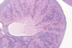
| MTB:18474 |
Leukocyte - Lymphocyte lymphoma |
Lymph node |
None (spontaneous) |
|
|
Female |
reproductive status not specified |
observed |
108 days |
Pancreatic lymph node, lymphoma. |
J:94314 |
|
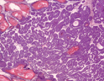
Image Caption:This high magnification allows to detail cytologic features of neoplastic cells. Neoplastic cells are closely-packed, round, with indistinct cell borders and a small amount of amphophilic cytoplasm. The nucleus is central, round, medium-sized and hypochromatic. Anisokaryosis and anisocytosis are mild. Mitotic index is high. Numerous neoplastic cells undergo single cell necrosis, giving a starry-sky appearance to the neoplasm.
|
|
|
Image ID:396 |
|
Source of Image:Mikaelian I |
|
Pathologist:Mikaelian I |
|
Method / Stain:H&E |
|
|
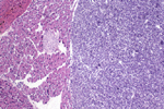
Image Caption:The pancreatic lymph node has been entirely replaced by a densely cellular neoplasm made of closely packed sheets of neoplastic lymphocytes. The pancreas is not infiltrated by neoplastic cells.
|
|
|
Image ID:395 |
|
Source of Image:Mikaelian I |
|
Pathologist:Mikaelian I |
|
Method / Stain:H&E |
|
|
|
| MTB ID |
Tumor Name |
Organ(s) Affected |
Treatment Type |
Agents |
Strain Name |
Strain Sex |
Reproductive Status |
Tumor Frequency |
Age at Necropsy |
Description |
Reference |
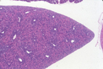
| MTB:18497 |
Leukocyte - Lymphocyte lymphoma |
Lung |
None (spontaneous) |
|
|
Female |
reproductive status not specified |
observed |
108 days |
Lung, lymphoma |
J:94314 |
|
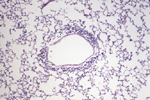
Image Caption:A few layers of neoplastic lymphocytes surround a vein.
|
|
|
Image ID:397 |
|
Source of Image:Mikaelian I |
|
Pathologist:Mikaelian I |
|
Method / Stain:H&E |
|
|
|
| MTB ID |
Tumor Name |
Organ(s) Affected |
Treatment Type |
Agents |
Strain Name |
Strain Sex |
Reproductive Status |
Tumor Frequency |
Age at Necropsy |
Description |
Reference |
| MTB:18498 |
Lung adenocarcinoma |
Lung |
None (spontaneous) |
|
|
Male |
reproductive status not specified |
observed |
229 days |
Lung, adenocarcinoma, bronchoalveolar |
J:94314 |
|
Image Caption:This higher magnification shows an area of continuity between the epithelial lining of a bronchus and the neoplasm. The neoplasm is made of tubules with an inconspicuous lumen. Neoplastic cells are medium-sized, with indistinct cell borders and a moderate amount of acidophilic cytoplasm. Their nucleus is central, round, and medium-sized. Anisokaryosis and anisocytosis are mild. There is a mild diffuse lymphocytic infiltration throughout the neoplasm.
|
|
|
Image ID:399 |
|
Source of Image:Mikaelian I |
|
Pathologist:Mikaelian I |
|
Method / Stain:H&E |
|
|
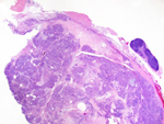
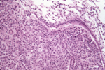
Image Caption:This is a densely cellular, unencapsulated, expensile, nodular neoplasm arising from the epithelium of a bronchus. This neoplasm is made of nests and tubules. It is associated with moderate atelectasis of the adjacent pulmonary parenchyma.
|
|
|
Image ID:398 |
|
Source of Image:Mikaelian I |
|
Pathologist:Mikaelian I |
|
Method / Stain:H&E |
|
|
|
| MTB ID |
Tumor Name |
Organ(s) Affected |
Treatment Type |
Agents |
Strain Name |
Strain Sex |
Reproductive Status |
Tumor Frequency |
Age at Necropsy |
Description |
Reference |
| MTB:18409 |
Mammary gland adenocarcinoma |
Mammary gland |
None (spontaneous) |
|
|
Female |
reproductive status not specified |
observed |
298 days |
Mammary gland, adenocarcinoma with epidermoid differentiation (adenoacanthoma) |
J:94314 |
|
Image Caption:The neoplasm is composed of interconnected trabeculae and nests. There is prominent squamous differentiation at the center of the largest nests. This specific type of mammary tumor is often refered to as "adenoacanthoma".
|
|
|
Image ID:387 |
|
Source of Image:Mikaelian I |
|
Pathologist:Mikaelian I |
|
Method / Stain:H&E |
|
|
|
| MTB ID |
Tumor Name |
Organ(s) Affected |
Treatment Type |
Agents |
Strain Name |
Strain Sex |
Reproductive Status |
Tumor Frequency |
Age at Necropsy |
Description |
Reference |
| MTB:18410 |
Mammary gland adenocarcinoma |
Mammary gland |
None (spontaneous) |
|
|
Female |
reproductive status not specified |
observed |
93 days |
Mammary gland, adenocarcinoma, glandular |
J:94314 |
|
Image Caption:This is a densely cellular neoplasm characterized by the formation of trabeculae and duct-like structures supported by a small to moderate amount of fibrovascular stroma.
|
|
|
Image ID:388 |
|
Source of Image:Mikaelian I |
|
Pathologist:Mikaelian I |
|
Method / Stain:H&E |
|
|
|
| MTB ID |
Tumor Name |
Organ(s) Affected |
Treatment Type |
Agents |
Strain Name |
Strain Sex |
Reproductive Status |
Tumor Frequency |
Age at Necropsy |
Description |
Reference |
| MTB:18472 |
Mammary gland adenocarcinoma |
Liver |
None (spontaneous) |
|
|
Female |
reproductive status not specified |
observed |
174 days |
Mammary adenocarcinoma, metastatic to the liver. |
J:94314 |
|
Image Caption:Liver: This higher magnification illustrates the margins of the metastatic mammary gland tumor. The metastasis is composed of tubules which are variably ectatic and are lined by a cuboidal epithelium. Some neoplastic tubules are ectatic and contain degenerated neutrophils. The center of the tumor has undergone extensive coagulation necrosis.
|
|
|
Image ID:393 |
|
Source of Image:Mikaelian I |
|
Pathologist:Mikaelian I |
|
Method / Stain:H&E |
|
|
Image Caption:Liver: a small metastasis of an mammary epithelial tumor is present in the subcapsular area. There is prominent coagulation necrosis at the center of the neoplasm
|
|
|
Image ID:392 |
|
Source of Image:Mikaelian I |
|
Pathologist:Mikaelian I |
|
Method / Stain:H&E |
|
|
|
| MTB ID |
Tumor Name |
Organ(s) Affected |
Treatment Type |
Agents |
Strain Name |
Strain Sex |
Reproductive Status |
Tumor Frequency |
Age at Necropsy |
Description |
Reference |
| MTB:27430 |
Muscle - Striated - Skeletal rhabdomyosarcoma |
Muscle - Striated - Skeletal |
None (spontaneous) |
|
|
Female |
reproductive status not specified |
observed |
474 days |
Muscle, striated, skeletal, thigh, rhabdomyosarcoma |
J:94314 |
|
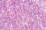
Image Caption:This is a densely cellular neoplasm composed of whorling and streaming bundles supported by a scant amount of fibrovascular stroma. Neoplastic cells are large, fusiform, with distinct cells borders and an abundant fibrillar acidophilic cytoplasm. They have 1-3 centrally located, medium-sized, ovale nuclei with a stippled chromatin. Anisocytosis is marked. Anisokaryosis is moderate.
|
|
|
Image ID:417 |
|
Source of Image:Mikaelian I |
|
Pathologist:Mikaelian I |
|
Method / Stain:H&E |
|
|
|
| MTB ID |
Tumor Name |
Organ(s) Affected |
Treatment Type |
Agents |
Strain Name |
Strain Sex |
Reproductive Status |
Tumor Frequency |
Age at Necropsy |
Description |
Reference |
| MTB:18501 |
Ovary adenoma |
Ovary |
None (spontaneous) |
|
|
Female |
reproductive status not specified |
observed |
469 days |
Ovary, granulosa cell tumor and papillary adenoma |
J:94314 |
|
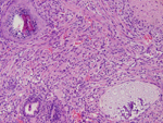
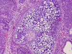
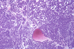
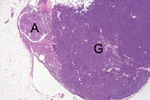
Image Caption:The ovary is entirely replaced by a large granulosa cell tumor (G) and a small papillary adenoma (A).
|
|
|
Image ID:418 |
|
Source of Image:Mikaelian I |
|
Pathologist:Mikaelian I |
|
Method / Stain:H&E |
|
|
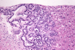
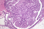
Image Caption:This is a high magnification of the ovarian papillary adenoma. It is composed of papillary projections and tubules separated by a moderate amount of fibrovascular stroma. The stroma contains numerous small nests of luteinized cells, a common feature of ovarian tubular adenomas.
|
|
|
Image ID:419 |
|
Source of Image:Mikaelian I |
|
Pathologist:Mikaelian I |
|
Method / Stain:H&E |
|
|
Image Caption:This is a high magnification of the ovarian papillary adenoma. The tumor is composed of a few tubules supported by a moderate to large amount of fibrovascular stroma. The tubules are lined by a one cell-thick low columnar epithelium. There is no evidence of atypia. The stroma is composed of numerous small nests of luteinized cells.
|
|
|
Image ID:420 |
|
Source of Image:Mikaelian I |
|
Pathologist:Mikaelian I |
|
Method / Stain:H&E |
|
|